
Home > News > Deciphering the Diagnostic Value of CD21: Illuminating Follicular Dendritic Cells in Lymphoid Tissue Lesions
Deciphering the Diagnostic Value of CD21: Illuminating Follicular Dendritic Cells in Lymphoid Tissue Lesions
- Concept
CD21, alternatively designated as the complement C3d receptor or type 2 complement receptor, is a pivotal cell surface glycoprotein. It fulfills dual critical roles: functioning as a key receptor within the complement cascade and serving as a cellular binding site for Epstein-Barr virus (EBV). Spatially, CD21 exhibits selective expression on follicular dendritic cells (FDCs), which form intricate reticular networks within the lymphoid follicle microenvironment. These networks are instrumental in antigen capture, retention, and presentation, while CD21-ligand interactions contribute to immune complex clearance and the regulation of B-cell activation and antibody responses.
Research Frontiers
Biological Traits and Functional Localization of CD21: Beyond its complement and EBV-binding functions, CD21’s restricted expression on FDCs underscores its significance as a specific marker for this cell population. FDCs, through CD21-mediated interactions, create a specialized niche that supports B-cell maturation, somatic hypermutation, and memory B-cell formation. This functional coupling positions CD21 as a linchpin in orchestrating adaptive immune responses within lymphoid tissues.
CD21 Expression in Reactive Lymphoid Hyperplasia: In lymphoid tissues undergoing reactive hyperplasia—an adaptive response to infection or inflammation—CD21 immunohistochemical staining reveals a highly organized, spherical reticular pattern. This structured network reflects the physiological arrangement of FDCs, which provide essential signals for B-cell activation, proliferation, and differentiation. The integrity of this CD21-positive reticulum serves as a hallmark of non-neoplastic lymphoid tissue, offering a critical morphological reference for diagnostic distinction.
Differential CD21 Expression in B-Cell Lymphomas: CD21 expression patterns exhibit substantial heterogeneity across B-cell lymphoma subtypes, enabling refined pathological classification. In follicular lymphoma and nodular lymphocyte-predominant Hodgkin lymphoma, CD21-positive FDCs form nodular proliferative structures that retain partial organizational features but diverge distinctly from reactive hyperplasia. In contrast, mantle cell lymphoma is characterized by sparse or scattered CD21-positive cells, a reflection of the disrupted tumor microenvironment. These subtype-specific expression profiles provide actionable diagnostic cues for pathologists.
Diagnostic Utility of CD21 in T-Cell Lymphomas: CD21 expression analysis also contributes significantly to T-cell lymphoma diagnosis. Angioimmunoblastic T-cell lymphoma exhibits a distinctive pattern wherein CD21-positive FDCs cluster around proliferating small blood vessels. Conversely, most peripheral T-cell lymphoma subtypes show either absent CD21 expression or a severely disrupted FDC network. These differences not only aid in distinguishing T-cell lymphoma subtypes but also offer insights into the underlying microenvironmental perturbations driving disease pathogenesis.
Specificity of CD21 in Follicular Dendritic Cell Tumors: CD21 demonstrates exceptional specificity as a diagnostic marker for follicular dendritic cell tumors (FDCTs), a rare group of mesenchymal neoplasms. FDCT cells consistently express CD21, enabling clear differentiation from other mesenchymal tumors. Immunohistochemical detection of CD21 thus provides definitive evidence for confirming the FDC origin of these tumors, supporting accurate diagnosis and classification. Notably, CD21 expression intensity and histological patterns may vary across FDCT subtypes, necessitating correlative analysis with other markers.
Technical Considerations and Interpretation Guidelines for CD21 Detection: Reliable CD21 immunohistochemical detection hinges on meticulous attention to technical variables, including sample preprocessing (e.g., antigen retrieval optimization), antibody dilution, and chromogenic system calibration. During result interpretation, emphasis must be placed not only on expression intensity but also on distribution pattern (e.g., reticular, nodular, scattered) and network integrity. To mitigate diagnostic bias, CD21 findings should be integrated with morphological features and other immunomarkers (e.g., CD20, CD3, CD10) in a comprehensive diagnostic algorithm.
Research Significance
CD21’s role as a specific FDC marker addresses a critical need in lymphoid tissue pathology: the accurate distinction between reactive and neoplastic lesions, and the subclassification of lymphomas. By delineating FDC network architecture and expression patterns, CD21 detection provides objective diagnostic evidence that enhances diagnostic precision, reduces interobserver variability, and guides clinical management. Furthermore, insights into CD21-mediated FDC functions advance our understanding of immune microenvironment dysregulation in lymphomas and autoimmune diseases, laying the groundwork for targeted therapeutic development.
Related Mechanisms, Research Methods, and Product Applications
Mechanisms
CD21 mediates its biological effects through dual pathways: as a complement receptor, it facilitates immune complex recognition and clearance; as an EBV receptor, it enables viral entry and contributes to virus-associated oncogenesis. On FDCs, CD21-dependent antigen presentation drives B-cell activation and antibody affinity maturation, while dysregulation of CD21 expression in neoplastic lesions reflects perturbations in immune microenvironment homeostasis.
Research Methods
Key research methods leveraging CD21 include:
* Immunohistochemistry (IHC) for in situ detection of CD21 expression and FDC network visualization in FFPE tissue samples.
* Flow cytometry for quantitative analysis of CD21 expression on B cells and FDCs in peripheral blood, bone marrow, or fresh tissue preparations.
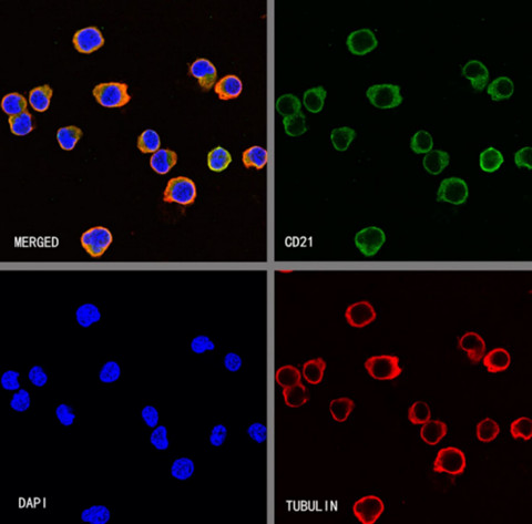
* Immunofluorescence co-staining to investigate CD21 colocalization with other immune markers (e.g., CD23, CD35) in lymphoid follicles.
* Functional assays (e.g., antigen presentation assays, B-cell proliferation assays) to elucidate CD21’s role in immune regulation.
Product Applications
ANT BIO PTE. LTD.’s CD21 antibodies, anchored by the STARTER brand’s "S-RMab® CD21 Recombinant Rabbit Monoclonal Antibody" (Catalog No.: S0B2043), are indispensable tools for lymphoid tissue research and clinical diagnosis:
* B-Cell Lymphoma Diagnosis & Classification: Enables accurate identification and subtyping of follicular lymphoma, chronic lymphocytic leukemia, and other B-cell malignancies by evaluating FDC network integrity.
* Immune Microenvironment & Germinal Center Research: Facilitates analysis of germinal center structure and function, and investigates FDC contributions to immune responses and disease pathogenesis.
* Autoimmune Disease Research: Supports studies on lymphoid follicle formation and functional status in conditions such as rheumatoid arthritis and systemic lupus erythematosus.
* Infection & Immunodeficiency Research: Aids in assessing lymphoid tissue structural abnormalities and immune dysfunction in immunocompromised patients.
The S0B2043 antibody, developed via ANT BIO PTE. LTD.’s proprietary S-RMab® recombinant rabbit monoclonal platform and validated for IHC, offers distinct advantages: high specificity with clear membrane localization (ensuring precise labeling of mature B cells and FDCs in FFPE samples) and exceptional staining stability with minimal batch variation—critical for consistent results in clinical diagnostics and translational research.
Brand Mission
ANT BIO PTE. LTD. is dedicated to empowering global life science research and clinical translation through the provision of high-quality, innovative biological reagents and solutions. Leveraging advanced development platforms—including recombinant rabbit monoclonal antibody, recombinant mouse monoclonal antibody, rapid monoclonal antibody, and multi-system recombinant protein expression platforms (E.coli, CHO, HEK293, Insect Cells)—coupled with rigorous certification (EU 98/79/EC, ISO9001, ISO13485), we strive to deliver reliable tools that accelerate scientific breakthroughs and improve patient outcomes.
Related Product List
| Catalog No. | Product Name | Host |
| S0B8377 | Alexa Fluor® 700 Mouse Anti-Human CD21 Antibody (S-764-18) | Mouse |
| S0B0178 | Alexa Fluor® 647 Mouse Anti-Human CD21 Antibody (S-764-18) | Mouse |
| S0B2043 | S-RMab® CD21 Recombinant Rabbit mAb (SDT-007-47) | Rabbit |
| S0B2043P | S-RMab® CD21 Recombinant Rabbit mAb, PBS Only (SDT-007-47) | Rabbit |
| S0B2003P | CD21 Recombinant Rabbit mAb, PBS Only (SDT-007-36) | Rabbit |
| S0B2003 | CD21 Recombinant Rabbit mAb (SDT-007-36) | Rabbit |
AI Disclaimer
This article is AI-compiled and interpreted based on the original work. All intellectual property (e.g., images, data) of the original publication shall belong to the journal and the research team. For any infringement, please contact us promptly and we will take immediate action.
ANT BIO PTE. LTD. – Empowering Scientific Breakthroughs
At ANTBIO, we are committed to advancing life science research through high-quality, reliable reagents and comprehensive solutions. Our specialized sub-brands (Absin, Starter, UA) cover a full spectrum of research needs, from general reagents and kits to antibodies and recombinant proteins. With a focus on innovation, quality, and customer-centricity, we strive to be your trusted partner in unlocking scientific mysteries and driving medical progress. Explore our product portfolio today and elevate your research to new heights.
Related News
- PTM Analysis is Essential for Antibody Research 7/10/2026
- BioMed X and Boehringer Ingelheim Expand XSeed Labs to Advance Next-Generation E 7/10/2026
- Everest Medicines Announces Close of Exclusive Licensing Agreement with Travere 7/10/2026
- Pan Modification Microspheres for PTM Proteomics 7/9/2026
- Specific Antibodies Drive Targeted Protein Degradation 7/9/2026
- Thermo Fisher Scientific Receives First FBI Approval for Rapid DNA Crime Scene P 7/9/2026
- Selective Chemical Labeling of Protein Lysine Methacrylation 7/8/2026
- Eppendorf Focuses Outreach Programmes to Advance STEM Education in Europe 7/8/2026
- Fumaroylation: A New Hub Linking Metabolism and Epigenetics 7/7/2026
- OECD Publishes Test Guideline for ToxTracker® 7/7/2026