
Reagents:
Molecular Biology
Biochemistry
Cell Biology
ELISA / Diagnostic Kits
Antibody
Serum/Medium
Other Reagents

- Human C5a ELISA Kit

- Product Detail
- Company Profile
Product Specification
| Usage | Required experimental equipment: 1. Microplate reader (450nm) 2. High-precision pipettes and pipette tips: 0.5-10uL, 5-50uL, 20-200uL, 200-1000uL 3. 37°C incubator 4. Distilled or deionized water Sample handling and requirements: The detection range of the kit is not equivalent to the concentration range of the analyte in the sample. Before the experiment, it is recommended to estimate the analyte concentration in the sample based on relevant literature and conduct preliminary experiments to determine the actual concentration in the sample. If the analyte concentration in the sample is too high or too low, dilute or concentrate the sample appropriately. If the sample type is not listed in the instructions, it is recommended to conduct a preliminary experiment to verify the validity of the test. Serum: Collect whole blood in a serum separator tube and place it at room temperature for 2 hours or at 2-8°C overnight. Then centrifuge at 1000×g for 20 minutes and remove the supernatant. Alternatively, store the supernatant at -20°C or -80°C, but avoid repeated freezing and thawing. Plasma: Collect the sample using EDTA or heparin as an anticoagulant. Within 30 minutes of collection, centrifuge at 1000×g for 15 minutes at 2-8°C. Remove the supernatant and test it. Alternatively, store the supernatant at -20°C or -80°C, but avoid repeated freezing and thawing. Tissue homogenate: Rinse the tissue with pre-chilled PBS (0.01M, pH 7.4) to remove any residual blood (lysed red blood cells in the homogenate may affect the test results). Weigh the tissue and mince it. Add the minced tissue to the appropriate volume of PBS (generally a 1:9 weight-to-volume ratio, e.g., 1g of tissue sample to 9mL of PBS. The specific volume can be adjusted according to experimental needs and recorded. It is recommended to add protease inhibitors to the PBS) in a glass homogenizer and grind thoroughly on ice or in a homogenizer. To further lyse tissue cells, the homogenate can be sonicated or repeatedly freeze-thawed. Finally, centrifuge the homogenate at 5000×g for 5-10 minutes and remove the supernatant for analysis. Cell culture supernatant: Centrifuge at 1000×g for 20 minutes. Remove the supernatant and analyze it immediately, or store it at -20°C or -80°C, but avoid repeated freeze-thaw cycles. Cell lysis buffer: Gently wash adherent cells with ice-cold PBS, then trypsinize and harvest the cells by centrifugation at 1000×g for 5 minutes. Suspension cells can be harvested directly by centrifugation. Wash the collected cells three times with pre-chilled PBS and resuspend in 150-200 μL of PBS per 1 × 10^6 cells (it is recommended to add protease inhibitors to the PBS; if the cell count is very low, reduce the PBS volume appropriately). Disrupt the cells by repeated freeze-thaw cycles or sonication. Centrifuge the extract at 1500 × g for 10 minutes at 2-8°C, and remove the supernatant for testing. Other biological fluids: Centrifuge at 1000 × g for 20 minutes, and remove the supernatant for testing. Sample Appearance: The sample should be clear and transparent, and any suspended matter should be removed by centrifugation. Sample Storage: Samples collected for testing within one week can be stored at 4°C. If testing is not possible, aliquot the sample into single-use aliquots and freeze at -20°C (for testing within one month) or -80°C (for testing within six months). Avoid repeated freeze-thaw cycles. Hemolysis of the sample can affect the final test results, so hemolyzed samples are not suitable for this test. Sample Dilution Scheme: Please estimate the sample concentration range in advance. If your test sample requires dilution, the reference dilution scheme is as follows: 100-fold dilution: One-step dilution. Add 5 μL of sample to 495 μL of universal diluent for a 100-fold dilution. 1000-fold dilution: Two-step dilution. Add 5 μL of sample to 95 μL of universal diluent for a 20-fold dilution. Then, add 5 μL of the 20-fold diluted sample to 245 μL of universal diluent for a 50-fold dilution, for a total of 1000-fold dilution. 100,000-fold dilution: Three-step dilution. Add 5 μL of sample to 195 μL of universal diluent and dilute it 40-fold. Then, add 5 μL of the 40-fold diluted sample to 245 μL of universal diluent and dilute it 50-fold. Finally, add 5 μL of the 2000-fold diluted sample to 245 μL of universal diluent and dilute it 50-fold, for a total dilution of 100,000-fold. For each dilution step, the sample volume should be at least 3 μL, and the dilution factor should not exceed 100-fold. Mix thoroughly after each dilution step to avoid foaming. Pre-test Preparation: 1. Remove the test kit from the refrigerator 10 minutes in advance and equilibrate to room temperature. 2. Prepare a gradient standard working solution: Add 1 mL of universal diluent to the lyophilized standard. Let stand for 15 minutes to completely dissolve, then gently mix (concentration 500 ng/mL). Then dilute to the following concentrations: 500 ng/mL, 250 ng/mL, 125 ng/mL, 62.5 ng/mL, 31.25 ng/mL, 15.62 ng/mL, 7.81 ng/mL, and 0 ng/mL. Serial dilution method: Take seven EP tubes and add 500 μL of universal diluent to each tube. Pipette 500 μL of the 500 ng/mL standard working solution into the first EP tube and mix thoroughly to make a 250 ng/mL standard working solution. Repeat this procedure for subsequent tubes. The last tube serves as a blank well; there is no need to pipette liquid from the penultimate tube. See the figure below for details. 3. Preparation of HRP Antibody Working Solution: 15 minutes before use, centrifuge 100 μL of concentrated HRP antibody at 1000 × g for 1 minute. Dilute the 100 μL concentrated HRP antibody to a 1 μL working concentration with universal diluent (e.g., 10 μL concentrate + 990 μL universal diluent). Prepare immediately before use. 4. Prepare 1×1 Wash Buffer: Dissolve 10 mL of 20×1 Wash Buffer in 190 mL of distilled water (Concentrated Wash Buffer removed from the refrigerator may crystallize, which is normal. Allow to stand at room temperature until the crystals have completely dissolved before preparing the buffer). Procedure: 1. Remove the desired strips from the aluminum foil bag after equilibration at room temperature for 10 minutes. Seal the remaining strips in a ziplock bag and return them to 4°C. 2. Sample Loading: Add 100 μL of sample or standard of varying concentrations to the corresponding wells. Add 100 μL of Universal Diluent to the blank wells. Cover with a film and incubate at 37°C for 60 minutes. (Recommendation: Dilute the samples to be tested at least 1-fold with Universal Diluent before loading into the plate. This minimizes the impact of matrix effects on the test results. When calculating sample concentrations, multiply by the corresponding dilution factor. It is recommended to run replicates for all samples and standards.) 3. Wash: Discard the liquid and add 300 μL of 1x wash buffer to each well. Let stand for 1 minute, shake off the wash buffer, and pat dry on absorbent paper. Repeat this process three times (a microplate washer can also be used). 4. Add HRP detection antibody: After washing, add 100 μL of HRP detection antibody working solution directly to each well. Cover with a film sealer and incubate at 37°C for 60 minutes. 5. Wash: Discard the liquid and wash the plate five times according to the procedure in step 3. 6. Add substrate: Add 90 μL of substrate (TMB) to each well, cover with a film sealer, and incubate at 37°C in the dark for 15 minutes. 7. Add stop solution: Remove the ELISA plate and add 50 μL of stop solution directly to each well. Immediately measure the OD value of each well at 450 nm. Calculation of Experimental Results: Result Interpretation: 1. Calculate the average OD value of the replicate wells containing the standard and sample and subtract the OD value of the blank well as the correction value. Plot a four-parameter logistic function standard curve on double-logarithmic graph paper, with concentration as the horizontal axis and OD value as the vertical axis. 2. If the sample OD value is higher than the upper limit of the standard curve, dilute the sample and retest it. Multiply the sample concentration by the corresponding dilution factor when calculating the sample concentration. 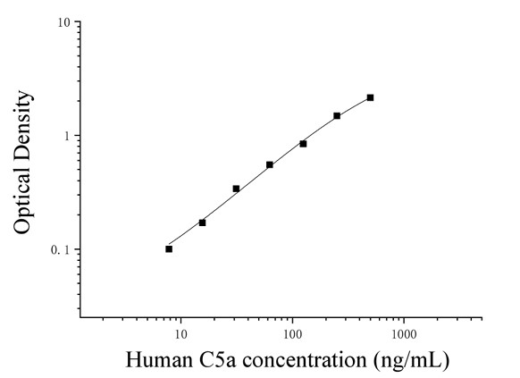 Note: This graph is for reference only. Sample content should be calculated using the standard curve plotted for each experimental data. Kit Performance: 1. Repeatability: Intra-plate coefficient of variation less than 10%, inter-plate coefficient of variation less than 10%. 2. Recovery: Human C5a was spiked into selected healthy human serum, plasma, and cell culture supernatant at three different concentrations, and the recovery was calculated. Note: This graph is for reference only. Sample content should be calculated using the standard curve plotted for each experimental data. Kit Performance: 1. Repeatability: Intra-plate coefficient of variation less than 10%, inter-plate coefficient of variation less than 10%. 2. Recovery: Human C5a was spiked into selected healthy human serum, plasma, and cell culture supernatant at three different concentrations, and the recovery was calculated.
Sensitivity | 3.8 ng/mL | Theory | This kit uses a double-antibody sandwich enzyme-linked immunosorbent assay (ELISA). Samples, standards, and HRP-labeled detection antibodies are sequentially added to microwells pre-coated with a capture antibody against human complement component 5a (C5a). After incubation and washing, the sample is developed using the substrate TMB. TMB converts to blue under the catalysis of HRP and to yellow under the action of acid. The intensity of the color is positively correlated with the amount of human complement component 5a (C5a) in the sample. The absorbance (OD) is measured at 450 nm using a microplate reader to calculate the sample concentration. | Source | Human | Synonym | Human Complement Component 5a ELISA Kit | Detection Type | Double antibody sandwich method | Description | Specificity: It can detect human C5a in samples and has no obvious cross-reaction with its analogs. | Composition |
Background | Complement component 5a (C5a) is a protein fragment released after the protease C5-convertase cleaves complement component C5 into C5a and C5b. C5a, another cleavage product of C5, is a highly inflammatory peptide that encourages complement activation, forms MACs, attracts innate immune cells, and releases histamine in allergic reactions. C5 originates in hepatocytes, but its synthesis can also be found in macrophages, where it may cause a local increase in C5a production. It is a chemoattractant and antiallergic agent; it is crucial in innate immunity but is also involved in adaptive immunity. Increased C5a production is associated with several inflammatory diseases. | General Notes | 1. Strictly adhere to the specified incubation time and temperature to ensure accurate results. All reagents must be at room temperature (20-25°C) before use. Refrigerate reagents immediately after use. | 2. Improper plate washing may result in inaccurate results. Ensure that all liquid in the wells is aspirated thoroughly before adding substrate. Do not allow the wells to dry out during incubation. 3. Remove any residual liquid and fingerprints from the bottom of the plate, as this will affect the OD value. 4. The substrate developer solution should be colorless or very light in color. Do not use substrate solution that has turned blue. 5. Avoid cross-contamination of reagents and specimens to prevent erroneous results. 6. Avoid direct exposure to strong light during storage and incubation. 7. Do not expose any reagents to bleaching solvents or the strong fumes emitted by bleaching solvents. Any bleaching agent will destroy the biological activity of the reagents in the kit. 8. Do not use expired products, and do not mix components with different product numbers and batches. 9. Recombinant proteins from sources other than the kit may not be compatible with the antibodies in this kit and will not be recognized. 10. If there is a possibility of disease transmission, all samples should be managed properly and samples and testing devices should be handled according to prescribed procedures. Storage Temp. | If the unopened kit is stored at 4°C, the shelf life is 6 months. | Test Range | 7.81-500 ng/mL | Applications | Serum, plasma, tissue homogenate, cell lysate, cell culture supernatant and other biological fluids |
|