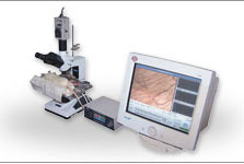
BI-2000 Medical Image Analysis System, developed in 1999 by Chengdu Tme Technology Co, Ltd, is a teaching and research oriental multi-function image analysis product. The design principal is providing more analysis functions with lower cost than other similar internal products.
The main characteristics of BI-2000 Medical Image Analysis System are Microcirculation Image and Physiology Parameter Integrated Observation, Dynamic Image Analysis, Digital Video and Analysis, Maze Automatic Tracking Analysis, Immune Tissue and Volume Calculation, Ion Channel Image Analysis, Static Image Processing and Analysis and Gel Electrophoresis Image Analysis etc.
After three-year function updating and customs’ feedback, more than fifty BI-2000 Medical Image Analysis Systems are assembled in national well-known medical schools, such as Peking Union Medical College, Tongji Medical University, West China Center of Medical Sciences and drug research institutes. Besides, several customs have already joined in the microcirculation laboratory for student experiments.
The hardware part of BI-2000 Medical Image Analysis System
1. Japanese JVC 480-wire Professional Color Camera, PAL compound video output
2. Thakit Professional Image Catching Card includs image catching, MPEG-1 digital recording and several function modules of television output.
3. XSZ-Hs7 second and first-class Triocular Biological Microscope made by Chongqing Optical Instrument Factory with a maximum magnify 1600.
4. Developing constant temperature experimental tables for rabbits, mice and frogs independently using together with microscopes.
5. Microcirculation Constant Temperature Control Device
6. Professional Direct Cameral Interface and Wide-vision Cameral Interface.
7. Zoom and Wide-angle Lens of 6mm-15mm.
8. Anti-pirating Dongle (Parallel Port)
9. Ten Times Optical Lens (It match up to Inverted Microscopes lengthen work distance)
The software part of BI-2000 Medical Image Analysis System
1. System Supporting Manual
2. The software disk of BI-2000 Medical Image Analysis System includes the following sixteen function modules.
1) Static Image Catching. 768x576 and 352x288 are changeable. Using standard image format: BMP non-packed and JPG packed format.
2) Digital Video Function supports VCD real-time recording.
3) Correcting the color of image in unison and printing multiple pictures. It is suitable for the printing of pictures of research papers.
4) Interactive Geometric Measurement (including measurements of straight line, curve, area, circumference).
5) Automatic Geometric Measurement Function.
6) Automatic counting of cells includes eliminating impurities, filling cavities, cutting apart targets, removing targets and improving the counting accuracy.
7) Dynamic Image Analysis Function and Digital Video Interactive Analysis can measure parameters of Change Margin, Velocity, Frequency etc, and be suitable for Pharmacological Analysis of Myocardial Cells.
8) Immunohistochemical Analysis automatically measuring parameters of Positive Distribution Area, Average Grey Scale, Average Optical Density and Integral Optical Density(IOD), and Supporting Grey Scale Section, Chromaticity Section and Hand Section.
9) Alignment Image Volume Measurement is used to the calculation of the volume of alignment section image and the area of body surface.
10) Microcirculation Image Experiments includes the comprehensive survey of indexes of image, electrocardiograph, blood pressure and breath, the measurement of blood diameter, blood speed and blood flow, the interactive measurement and recording of fifteen experimental parameters, the recording and replay of image and physiological waves, and the printing of experimental picture and text report. Microcirculation Image Observation Object Lens is classified by four times, ten times and forty times. The clearness is much better than similar internal products.
11) Ion Channel Image Analysis is used to analysis ion channel output waves and measure the open and close time of the channel.
12) Water-maze Tracking Analysis Software is used to recognize targets automatically, follow target tracking and count time, distance, and the efficiency of six periods in quadrant and ring methods.
13) Dynamic Tracking Analysis Software is used to trail activity time and rout of most sixteen animals in each activity area and the pharmacological experiments reflecting the animal activity at the same time automatically.
14) Gel Image Analysis Software is used to analysis electrophoresis strip image automatically reaching Molecular Weight, Strip Displacement, Strip Integral Optical Density (IOD) and the Concentration Percent of each strip in swimming lane. It can also be used to analysis Spot Hybrid Images and results of Integral Optical Density (IOD) and Mass of each spot.
15) Electron Case Image Database Management Software supports the preservation of patient and diagnoses information, fuzzy enquiry, modification, elimination and printing. There are most four images in each case.
16) Multimedia Teaching Management function includes Digital Recording Teaching, Teaching Section Image Database and supports Teaching Section Instruction and Image Comparison Magnify function.
Characteristic
1. Complete microcirculation observation and analysis experimental platform.
2. Immunohistochemical Analysis, dynamic and static image measurement.
3. As many as fifteen software function models satisfy the need of research and teaching.


