
Reagents:
Molecular Biology
Biochemistry
Cell Biology
ELISA / Diagnostic Kits
Antibody
Serum/Medium
Other Reagents

- Human IL-8/CXCL8 ELISA Kit

- Product Detail
- Company Profile
Product Specification
| Usage | Need to bring your own test equipment 1. Microplate reader (can measure the absorption value of 450nm detection wavelength and the absorption value of 540nm or 570nm correction wavelength) 2. High-precision liquid dispenser and disposable tip 3. Distilled water or deionized water 4. Bottle washing (spray bottle), multi-channel plate washer or automatic plate washer 5. 500mL measuring cylinder 1. Preparation before the experiment 1. Sample collection and storage ① Cell culture supernatant: particulate matter should be removed by centrifugation; Test the sample immediately. If the sample is not tested in time after collection, it is recommended to pack it according to the one-time usage amount and store it in a refrigerator at-20 ℃ to avoid repeated freezing and thawing. Samples may need to be diluted with diluent (1 ×). ② Serum: Use a serum separation tube (SST) to collect samples, and place the samples at room temperature for 30 minutes. Centrifuge for 15 minutes at a rotation speed of 1000 g. The serum was removed immediately and tested immediately. If the sample is not tested in time after collection, it is recommended to pack it according to the one-time usage amount and store it in a refrigerator ≤-20 ℃ to avoid repeated freezing and thawing. Samples may need to be diluted with diluent (1 ×). ③ Plasma: Plasma was collected using EDTA, heparin or citric acid as anticoagulant, centrifuged for 15 minutes within 30 minutes after collection, rotated at 1000g, and detected immediately. If the sample is not tested in time after collection, it is recommended to pack it according to the one-time usage amount and store it in a refrigerator ≤-20 ℃ to avoid repeated freezing and thawing. Samples may need to be diluted with diluent (1 ×). 2. Reagent preparation (Please place all reagents and samples at room temperature before use and let them stand15Minutes. All experimental samples and standards are recommendedDo repeat hole detection) ① Preparation of 1 × washing liquid: The concentrated washing liquid in the kit is 20 × mother liquid, which needs to be diluted into 1 × working liquid with distilled water before use.Example:Take 10mL of concentrated washing solution + 190mL of distilled water and make the volume to 200mL. In actual operation, the amount used can be calculated first, and then prepared. ② Preparation of 1 × dilution buffer: The concentration and dilution buffer in the kit is 10 × mother liquor, which should be diluted to 1 × working solution with distilled water before use.Example:Make up to 30 mL with 3 mL of concentration and dilution buffer + 27 mL of distilled water. In actual operation, the required amount of dilution buffer solution can be calculated according to the sample dilution factor, and then prepared. ③ Antibody detection: centrifuge the dry powder to the bottom of the tube, dissolve it with 110uL dilution buffer (1 ×), and let it stand at room temperature for 5 minutes to obtain 100 × mother liquor; Dilute to 1 × working solution before use. Calculate the required volume according to the dosage of 100uL per well.Example:After 10 wells were used, 10 uL of the detection antibody having a working concentration of 100 times was taken, and the volume was diluted to 1 mL using a dilution buffer (1 ×) to obtain 1 mL of the detection antibody having a working concentration of 1 ×. ④ SA-HRP: SA-HRP is 40 × mother liquor, which needs to be diluted with dilution buffer (1 ×) before use to prepare 1 × working solution, and the required amount per well is 100uL.Example:After 10 wells were used, 25 uL of 40 × mother liquor + 975 uL of dilution buffer (1 ×) was diluted to 1 mL to obtain 1 mL of detection antibody having a 1 × working concentration. ⑤ Color development solution: According to 100uL per well, calculate the dosage required for the current test, take out the corresponding volume of color development solution, and protect it from light; The chromogenic solution removed is for the same day only. ⑥ Standard: The freeze-dried standard is re-dissolved with dilution buffer (1 ×), and the re-dissolving volume is 1000uL to obtain the standard mother liquor with a concentration of 2000pg/mL. Gently shake for at least 5 minutes and it dissolves well. 300 uL of dilution buffer (1 ×) was added to each dilution tube. Make serial dilutions of the standard mother liquor according to the figure below, and each tube must be thoroughly mixed before pipetting to the next tube. The standard mother solution without dilution can be used as the highest point of the standard curve (2000 pg/mL), and the dilution buffer (1 ×) can be used as the zero point of the standard curve (0 pg/mL). 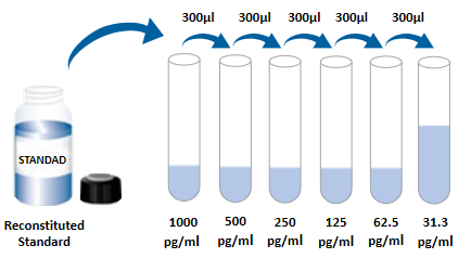 2. Operation steps 1. Prepare all required reagents and standards; 2. Take out the microplate from the sealed bag that has been balanced to room temperature. Please put the unused slats back into the aluminum foil bag and reseal them; 3. Add 300uL of washing liquid to the microplate, let it stand and soak for 30 seconds, discard the washing liquid and pat the microplate dry on absorbent paper. Please use it immediately and do not let the microplate dry; 4. Add different concentration standards, experimental samples or quality control products to the corresponding wells, 100uL per well. Sealing the reaction hole with plate sealing adhesive paper and incubating at room temperature for 2 hours; 5. Suck off the liquid in the plate and wash the plate with a bottle washer, a multi-channel plate washer or an automatic plate washer. 300 uL of washing solution was added to each well, and then the washing solution in the plate was aspirated off. Repeat the operation 3 times. Trying to absorb the residual liquid as much as possible every time you wash the plate will help to get good experimental results. At the end of the last plate washing, please suck all the liquid in the plate or turn the plate upside down, and pat all the residual liquid dry on absorbent paper; 6. Add 100 uL of detection antibody to each well. Seal the reaction wells with plate sealing tape, and incubate at room temperature for 2 hours; 7. Repeat the plate washing operation in step 5; 8. Add 100uLSA-HRP to each well and incubate at room temperature for 20 minutes. Be careful to avoid light; 9. Repeat the plate washing operation in step 5; 10. Add 100uL of chromogenic solution to each microwell, incubate at room temperature for 5-30 minutes, and avoid light; 11. Add 50uL of stop solution to each microwell, and the color of the solution in the well will change from blue to yellow. If the color of the solution changes to green or the color changes are inconsistent, pat the microplate gently to mix the solution evenly; 12. Within 30 minutes after adding the stop solution, measure the absorbance value of 450nm using a microplate reader, and set 540nm or 570nm as the calibration wavelength. If dual-wavelength correction is not used, the accuracy of the results may be affected; 13. Calculation Results: Average the corrected absorbance values (OD450-OD540 or OD570), multiple well readings for each standard and sample, and then subtract the average zero standard OD value. Four-parameter logic (4-PL) curve fitting was performed using computer software to create the standard curve. Alternatively, a curve can be generated by plotting the logarithm of the standard concentration versus the logarithm of the corresponding OD value, and the best fit line can be determined by regression analysis. This process can generate a data fit that is sufficiently useful but less accurate. If the sample is diluted, the concentration should be calculated by multiplying the dilution factor.  3. Kit parameters 1. Recovery: Different levels of human IL-8/CXCL8 were spiked into cell culture medium samples, and the recovery rate was determined. The recoveries ranged from 96 to 118%, with an average recovery of 108%. 2. Sensitivity: The lowest measurable dose (MDD) of human IL-8/CXCL8 is generally less than 7.8 pg/mL. The lowest measurable value is the corresponding concentration calculated from the mean of the zero-point absorbance values of 20 standard curves plus two standard deviations. 3. Calibration: This ELISA kit was calibrated with high purity recombinant human IL-8/CXCL8 protein expressed by E. coli. 4. Linearity: 4 different samples were spiked with high concentrations of human IL-8/CXCL8, and then the samples were diluted to the detection range with diluent (1 ×) to determine their linearity.
4. Analysis of frequently asked questions 1. Whiteboard (after the color development is completed, no color appears)
| |||||||||||||||||||||||||||||||||||||||||||||||||||||||||||||||||||||||||||||||
| Species Reactivity | Human | |||||||||||||||||||||||||||||||||||||||||||||||||||||||||||||||||||||||||||||||
| Theory | This kit adopts double antibody sandwich enzyme-linked immunosorbent detection technology. Specific anti-human IL-8 antibodies were pre-coated on high affinity plates. The standard substance, the sample to be tested and the biotinylated detection antibody are added to the well of the enzyme label plate, and after incubation, IL-8 present in the sample binds to the solid phase antibody and the detection antibody to form an immune complex. After washing to remove unbound material, horseradish peroxidase-labeled Streptavidin-HRP was added. After washing, a chromogenic substrate is added to protect the color from light. A stop solution was added to stop the reaction, and the absorbance value was measured at a wavelength of 450 nm (reference calibration wavelength of 540 nm or 570 nm). | |||||||||||||||||||||||||||||||||||||||||||||||||||||||||||||||||||||||||||||||
| Synonym | 3-10C, AMCF-I, C-X-C motif chemokine 8, CXCL8, CXCL8SCYB8, Emoctakin, GCP1, GCP-1TSG-1, IL8, IL-8, interleukin 8, K60, LAI, LECT, MDNCF, MDNCFb-ENAP, member 8, MONAPGCP1, NAP1, NAP-1NAP1, NCF, Neutrophil-activating protein 1, Protein 3-10C, T cell chemotactic factor, T-cell chemotactic factor, TCF, TSG1 | |||||||||||||||||||||||||||||||||||||||||||||||||||||||||||||||||||||||||||||||
| Composition | Please use it within the validity period of the kit (new and old products are shipped randomly)
| |||||||||||||||||||||||||||||||||||||||||||||||||||||||||||||||||||||||||||||||
| Background | Interleukin-8 (IL-8, also known as GCP-1, NAP-1, and CXCL8) is an 8-9kDa chemokine belonging to the α-type or CXC family that binds to heparin. So far, a total of 15 human CXC family proteins have been found, ranging in size from 8 to 12kDa. Most of them are located on human chromosome 4, all have a typical triple β-sheet layer/an α-helix structure, and most of them show Glu-Leu-Arg tripeptide motif at the N-terminus. Human IL-8 was first synthesized as a 99 amino acid precursor containing a 20 amino acid signal sequence and a 79 amino acid mature region. It can be circulated in vivo as monomers, homodimers, and heterodimers formed with CXCL4/PF4. IL-8 monomers are considered to be the most biologically active, whereas heterodimers may enhance the activity of PF4. At the amino acid level, mature human IL-8 has 65% and 70% homology to pigs and dogs, respectively. In rodents, the corresponding gene for IL-8 has not been found. A variety of IL-8 isoforms were generated by alternative splicing and differential proteolysis. Alternative splicing occurs at the C-terminus, where there is an 11 amino acid substitution (at aa # 92-99). Protease hydrolysis may be a cell-specific event that produces a truncation at the N-terminus of IL-8. For example, fibroblasts and vascular endothelial cells cleave amino acids 21 and 22 to form IL-8, while monocytes and lymphocytes cleave amino acids 21-25 to produce IL-8. These short IL-8s generally have higher biological activity, especially against the CXCR1IL-8 receptor. Approximately 15% of IL-8 is citrullinated at the precursor Arg27 position, which increases the half-life of IL-8 and promotes leukocytosis. Many types of cells secrete IL-8, including monocytes and neutrophils, fibroblasts and keratinocytes, mast cells, visceral smooth muscle cells, dendritic cells, type II large alveolar cells, and endothelial cells. There are two IL-8 receptors, both of which belong to G protein-coupled receptor proteins: CXCR1/IL-8RA and CXCR2/IL-8RB, and the amino acid sequences between them share 77% homology. CXCR1 has a molecular weight of 45-50 kDa and is almost entirely exclusive to IL-8; CXCR2 has a molecular weight of 35-40 kDa and is common to all CXC chemokines. CXCR1 and CXCR2, respectively, form constitutive homodimers, which appear to be their functional configuration. Heterodimers also form when expressed in the same cell; But when it binds to IL-8, it will be disintegrated. CXCR2 responds to low concentrations of IL-8 and is mainly related to chemotaxis and MMP-9 release. In contrast, CXCR1 responds to high concentrations of IL-8 and is associated with respiratory bursts and activation of phospholipase D2. Therefore, in neutrophils CXCR2 is thought to direct the migration of neutrophils to the site of inflammation and then elicit CXCR1-mediated antibacterial activity. The best known function of IL-8 is its pro-inflammatory effect in immune cells. Essentially, IL-8 is secreted by a variety of cell types exposed to inflammatory stimulatory factors. For monocytes/macrophages, microbial exposure causes the release of IL-8; Subsequently, CXCR2-mediated chemotaxis migrates neutrophils to the site of antigen attack, accompanied by the activation and initiation of antibacterial activity in the next step. IL-8 can enhance the effect of M-CSF in bone marrow, thereby causing the maturation and release of granulocytes. These two functions of IL-8 complement each other. It has been reported that IL-8 has angiogenic function on tumor-associated endothelial cells. At this time, tumor-derived IL-8 can activate CXCR1 and CXCR2 in vascular endothelial cells in a paracrine manner. Both CXCR1 and CXCR2 are associated with the PI3-K/Akt and RasGTP signaling pathways and are involved in cell survival and proliferation. In addition, IL-8 positively regulates VEGFR2 and EGFR, which belong to receptor tyrosine kinases that mediate cell growth and migration. | |||||||||||||||||||||||||||||||||||||||||||||||||||||||||||||||||||||||||||||||
| General Notes | 1. Please use the kit within the validity period. 2. The components of different kits and different batch kits cannot be mixed. 3. If the sample value is greater than the highest value of the standard curve, the sample should be diluted with diluent (1 ×) and re-tested; If the cell culture supernatant sample needs to be distributed and diluted, cell culture medium can be used for other intermediate dilutions except dilution with diluent in the last step. 4. Differences in test results can be caused by a variety of factors, including the operation of the experimenter, the use of the pipette, the plate washing technique, the reaction time or temperature, the storage of the kit, etc. 5. The terminating solution in the kit is an acidic solution. Please protect your glasses, hands, face and clothes when using it. 6. For scientific research only, not for in vitro diagnosis. | |||||||||||||||||||||||||||||||||||||||||||||||||||||||||||||||||||||||||||||||
| Storage Temp. | Kit unopened, stored at 2-8 ℃. | |||||||||||||||||||||||||||||||||||||||||||||||||||||||||||||||||||||||||||||||
| Test Range | 31.3pg/mL-2000pg/mL |
-
AntBio is a biotechnology group company dedicated to serving life sciences, aiming to help scientists accelerate research and improve work efficiency. AntBio provides comprehensive and high-quality reagent tools for basic research, drug development, and diagnosis, including research grade antibodies, proteins, biochemical reagents, and assay kits. These research tools are widely used in different segments of life science research. The group company currently consists of three brands, Absin, Starter-Bio and UA-Bio.
| Request Information |
| Other Products |
| Related Products |
| Recently viewed products |