

- Human IL-23 ELISA Kit

- Product Detail
- Company Profile
Product Specification
| Usage | Self-prepared test equipment required for the experiment Microplate reader (can measure the absorption value of 450nm detection wavelength and the absorption value of 540nm or 570nm correction wavelength) High precision liquid dispenser and disposable tip Distilled or deionized water Bottle washer (spray bottle), multi-channel plate washer or automatic plate washer 500mL measuring cylinder 1. Preparation before the experiment (1) Sample collection and storage:Cell culture supernatant: particulate matter should be removed by centrifugation; Test the sample immediately. If the sample is not tested in time after collection, it is recommended to pack it according to the one-time usage amount and store it in a refrigerator at-20 ℃ to avoid repeated freezing and thawing. Samples may need to be diluted with diluent (1 ×). Serum: Samples were collected using a serum separation tube (SST) and the samples were left at room temperature for 30 minutes. Centrifuge for 15 minutes at 1000 g. The serum was removed immediately and tested immediately. If the sample is not tested in time after collection, it is recommended to pack it according to the one-time usage amount and store it in a refrigerator ≤-20 ℃ to avoid repeated freezing and thawing. Samples may need to be diluted with diluent (1 ×). Plasma: Plasma was collected using EDTA, heparin, or citric acid as anticoagulant, centrifuged for 15 min within 30 min after collection at a rotation speed of 1000 g, and tested immediately. If the sample is not tested in time after collection, it is recommended to pack it according to the one-time usage amount and store it in a refrigerator ≤-20 ℃ to avoid repeated freezing and thawing. Samples may need to be diluted with diluent (1 ×). (2) Preparation work before testing: Please place all reagents and samples at room temperature for 15 minutes before use. It is recommended that all experimental samples and standards be repeatedly tested 1 × Wash solution preparation:The concentrated washing liquid in the kit is 20 × mother liquid, which needs to be diluted into 1 × working liquid with distilled water before use. Example: Take 10ml of concentrated washing liquid + 190mL of distilled water and make the volume to 200mL. In actual operation, the amount used can be calculated first, and then prepared. 1 × Buffer preparation for dilution:The buffer solution for concentration and dilution in the kit is 10 × mother liquor, which should be diluted to 1 × working solution with distilled water before use. Example: Take 3ml of concentration and dilution buffer + 27mL of distilled water and make the volume to 30mL. In actual operation, the required amount of dilution buffer solution can be calculated according to the sample dilution factor, and then prepared. Antibody detected:Centrifuge the dry powder to the bottom of the tube, dissolve it with 110uL of dilution buffer (1 ×), and let it stand at room temperature for 5 minutes to obtain 100 × mother liquor; Dilute to 1 × working solution before use. Calculate the required volume according to the dosage of 100uL per well. Example: Ten wells are used, and 10 uL of the detection antibody having a 100-fold working concentration is taken, and the volume is diluted to 1 mL using a dilution buffer (1 ×) to obtain 1 ml of the detection antibody having a 1 × working concentration. SA HRP:SA-HRP is 40 × mother solution. Before use, it needs to be diluted with dilution buffer (1 ×) to prepare 1 × working solution. The required amount per well is 100 uL. Example: After 10 wells are used, 25 uL of 40 × mother liquor + 975 uL of dilution buffer (1 ×) is taken and diluted to 1 mL to obtain 1 ml of 1 × working concentration detection antibody. Color developer:According to 100 uL per well, calculate the dosage required for the current test, take out the corresponding volume of chromogenic agent, and protect it from light; The developer removed is for the same day use only. Standard:The lyophilized standard was redissolved with dilution buffer (1 ×), and the redissolved volume was 1000uL to obtain a standard mother liquor with a concentration of 8000pg/mL. Gently shake for at least 5 minutes and it dissolves well. 300 uL of dilution buffer (1 ×) was added to each dilution tube. Make serial dilutions of the standard mother liquor according to the figure below, and each tube must be thoroughly mixed before pipetting to the next tube. The standard mother solution without dilution can be used as the highest point of the standard curve (8000 pg/mL), and the dilution buffer (1 ×) can be used as the zero point of the standard curve (0 pg/mL). 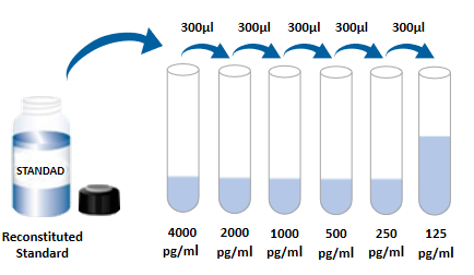 (1) Prepare all required reagents and standards; (2) Take out the microplate from the sealed bag that has been balanced to room temperature. Please put the unused slats back into the aluminum foil bag and reseal them; (3) Add 300uL of washing solution to the microplate, let it stand and soak for 30 seconds, discard the washing solution and pat the microplate dry on absorbent paper. Please use it immediately and do not let the microplate dry; (4) Add different concentrations of standards, experimental samples or quality controls to the corresponding wells, 100 uL per well. Sealing the reaction hole with plate sealing adhesive paper and incubating at room temperature for 2 hours; (5) Suck off the liquid in the plate, and wash the plate with a bottle washer, a multi-channel plate washer or an automatic plate washer. 300 uL of washing solution was added to each well, and then the washing solution in the plate was aspirated off. Repeat the operation 3 times. Trying to absorb the residual liquid as much as possible every time you wash the plate will help to get good experimental results. At the end of the last plate washing, please suck all the liquid in the plate or turn the plate upside down, and pat all the residual liquid dry on absorbent paper; (6) Add 100 uL of detection antibody to each microwell. Seal the reaction wells with plate sealing tape and incubate at room temperature for 2 hours; (7) Repeat the plate washing operation in step 5; (8) 100 uL of SA-HRP was added to each well and incubated at room temperature for 20 minutes. Be careful to avoid light; (9) Repeat the plate washing operation in step 5; (10) Add 100 uL of chromogenic solution to each microwell, incubate at room temperature for 5-30 minutes, and pay attention to protecting from light; (11) Add 50 uL of stop solution to each well and the color of the solution in the well will change from blue to yellow. If the color of the solution changes to green or the color changes are inconsistent, pat the microplate gently to mix the solution evenly; (12) Within 30 minutes after addition of the stop solution, the absorbance value at 450 nm was measured using a microplate reader, and 540 nm or 570 nm was set as the correction wavelength. If dual-wavelength correction is not used, the accuracy of the results may be affected; (13) Calculation results: Average the corrected absorbance values (OD450-OD540/OD570) and multiple well readings of each standard and sample, and then subtract the average zero standard OD value. Four-parameter logic (4-PL) curve fitting was performed using computer software to create the standard curve. Alternatively, a curve can be generated by plotting the logarithm of the standard concentration versus the logarithm of the corresponding OD value, and the best fit line can be determined by regression analysis. This process can generate a data fit that is sufficiently useful but less accurate. If the sample is diluted, the concentration should be calculated by multiplying the dilution factor.
5. specificity This ELISA can detect native and recombinant human IL-23 protein. The following factors were formulated with diluent (1 ×) at a concentration of 50 ng/mL to detect cross-reactivity with human IL-23. Interference with human EGF was detected by incorporating 50 ng/mL of the interfering factor into the mid-range recombinant human IL-23 control. No significant cross-reactivity or interference was observed. Recombinant human IL-12 Rβ There was no cross-reaction between 1/Fc Chimera and recombinant human IL-23R/Fc Chimera, but there was interference at concentrations > 3125 pg/mL.
4. Analysis of frequently asked questions
2. Flower plate (blank and negative positive controls are normal, but the OD value of the specimen well is obviously higher)
| |||||||||||||||||||||||||||||||||||||||||||||||||||||||||||||||||||||||||||
| Species Reactivity | Human | |||||||||||||||||||||||||||||||||||||||||||||||||||||||||||||||||||||||||||
| Theory | This kit adopts double antibody sandwich enzyme-linked immunosorbent detection technology. Specific anti-human IL-23 antibodies were pre-coated on high affinity plates. The standard substance, the sample to be tested and the biotinylated detection antibody are added to the well of the enzyme label plate, and after incubation, IL-23 present in the sample binds to the solid phase antibody and the detection antibody to form an immune complex. After washing to remove unbound material, horseradish peroxidase-labeled Streptavidin-HRP was added. After washing, a chromogenic substrate is added to protect the color from light. A stop solution was added to stop the reaction, and the absorbance value was measured at a wavelength of 450 nm (reference calibration wavelength of 540 nm or 570 nm). | |||||||||||||||||||||||||||||||||||||||||||||||||||||||||||||||||||||||||||
| Synonym | IL-23 p19/IL-12 p40, IL23, IL-23, IL-23A, IL-23-A, IL-23p19, IL-23p19/IL-12p40, IL23P19P19, interleukin 23 p19 subunit, interleukin 23, alpha subunit p19, interleukin-23 subunit alpha, Interleukin-23 subunit p19, JKA3 induced upon T-cell activation, MGC79388, SGRF, SGRFIL-23 subunit alpha | |||||||||||||||||||||||||||||||||||||||||||||||||||||||||||||||||||||||||||
| Detection Type | Double antibody sandwich method | |||||||||||||||||||||||||||||||||||||||||||||||||||||||||||||||||||||||||||
| Composition | Please use within the expiration date of the kit
| |||||||||||||||||||||||||||||||||||||||||||||||||||||||||||||||||||||||||||
| Background | Interleukin 23 (IL-23) is a disulfide-linked cytokine consisting of the p19 subunit specific to IL-23 and the p40 subunit shared by IL-12. It is produced by activated macrophage, microglia, and monocyte-derived dendritic cells in response to attack by pathogens including certain bacteria and viruses and/or their components. The functional IL-23 receptor complex comprises two receptor subunits, an IL-12 receptor β1 subunit (IL-12Rβ1) and an IL-23-specific receptor subunit (IL-23R). The receptor complex is expressed in mouse Th1, Th2 cells, bone marrow dendritic cells, activated macrophages and CD4+CD45Rb (low) memory T cells. The IL-23 immune pathway induces the earliest neutrophil recruitment to the foci of infection. IL-23 also enhances the development and maintenance of Th17 cells. It promotes Th17-mediated autoimmunity and tumor progression. In humans, IL-23 expression is upregulated in several immune dysfunctional diseases including psoriasis, Crohn's disease, and multiple sclerosis. | |||||||||||||||||||||||||||||||||||||||||||||||||||||||||||||||||||||||||||
| General Notes | (1) For scientific research only, not for in vitro diagnosis; (2) Please use it within the validity period of the kit; (3) The components of different kits and kits with different batch numbers cannot be mixed; (4) If the sample value is greater than the highest value of the standard curve, the sample should be diluted with diluent (1 ×) and re-tested; If the cell culture supernatant sample needs to be distributed and diluted, cell culture medium can be used for other intermediate dilutions except for the last step of dilution with diluent; (5) Differences in test results can be caused by a variety of factors, including the operation of the experimenter, the use of the pipette, the plate washing technique, the reaction time or temperature, the storage of the kit, etc. (6) The terminating solution in the kit is acidic solution. Please protect your glasses, hands, face and clothes when using it. | |||||||||||||||||||||||||||||||||||||||||||||||||||||||||||||||||||||||||||
| Storage Temp. | Kit unopened, stored at 2-8 °C. | |||||||||||||||||||||||||||||||||||||||||||||||||||||||||||||||||||||||||||
| Test Range | 125.0 - 8,000 pg/mL |
 Note:The standard curve data provided is for reference only, and the sample content should be calculated according to the standard curve drawn in the same test.
-
AntBio is a biotechnology group company dedicated to serving life sciences, aiming to help scientists accelerate research and improve work efficiency. AntBio provides comprehensive and high-quality reagent tools for basic research, drug development, and diagnosis, including research grade antibodies, proteins, biochemical reagents, and assay kits. These research tools are widely used in different segments of life science research. The group company currently consists of three brands, Absin, Starter-Bio and UA-Bio.
| Request Information |
| Other Products |
| Related Products |