| Usage | Sample Collection, Processing, and Storage Methods: 1. Serum: Use pyrogen- and endotoxin-free tubes. Avoid any cell stimulation during handling. After blood collection, centrifuge at 3000 rpm for 10 minutes to quickly and carefully separate the serum from the red blood cells. 2. Plasma: Anticoagulate with EDTA, citrate, or heparin. Centrifuge at 3000 rpm for 30 minutes and remove the supernatant. 3. Cell Supernatant: Centrifuge at 3000 rpm for 10 minutes to remove particles and aggregates. 4. Tissue Homogenate: Add an appropriate amount of saline to the tissue and mash. Centrifuge at 3000 rpm for 10 minutes and remove the supernatant. 5. Storage: If samples are not tested immediately after collection, aliquot them into single-use aliquots and freeze at -20°C. Avoid repeated freeze-thaw cycles. Thaw at room temperature and ensure that the sample is evenly and thoroughly thawed. Supplies You Need
1. Microplate reader (450nm)
2. High-precision pipettes and tips: 0.5-10µL, 2-20µL, 20-200µL, 200-1000µL
3. 37°C incubator
Reagent Preparation:
20×Wash Buffer: Dilute 1:20 with distilled water, i.e., add 1 part 20×Wash Buffer to 19 parts distilled water.
Plate Washing Method:
1. Manual Plate Washing: Shake off all liquid from the wells, fill each well with wash buffer, let stand for 1 minute, shake off all liquid from the wells, pat dry on absorbent paper, and wash the plate five times.
2. Automatic Plate Washer: Add 350µL of wash buffer to each well, soak for 1 minute, and wash the plate five times.
Procedure:
1. Remove the desired strips from the aluminum foil bag after equilibration at room temperature for 20 minutes. Seal the remaining strips in a ziplock bag and return them to 4°C.
2. Arrange the standard and sample wells. Add 50 μL of the standard of different concentrations to each standard well.
3. Add 10 μL of the sample to be tested to the sample wells, followed by 40 μL of sample diluent. Leave blank wells untouched.
4. Add 100 μL of horseradish peroxidase (HRP)-conjugated detection antibody to each standard and sample well, except for the blank wells. Seal the wells with sealing film and incubate at 37°C in a water bath or incubator for 60 minutes.
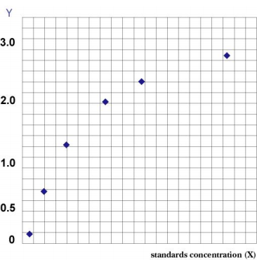
5. Discard the liquid, pat dry on absorbent paper, fill each well with wash solution, let stand for 1 minute, shake off the wash solution, and pat dry on absorbent paper. Repeat this process five times (a plate washer can also be used). 6. Add 50 μL each of substrates A and B to each well and incubate at 37°C in the dark for 15 minutes. 7. Add 50 μL of stop solution to each well and measure the OD value of each well at a wavelength of 450 nm within 15 minutes. Result Interpretation: Draw a standard curve: In an Excel worksheet, plot the standard concentration on the horizontal axis and the corresponding OD value on the vertical axis. Calculate the concentration of each sample using the curve equation.
Kit Performance
1. Accuracy: The correlation coefficient (R) between the linear regression of the standard and the expected concentration is greater than or equal to 0.9900.
2. Sensitivity: The minimum detectable concentration is less than 0.1 μg/mL.
3. Specificity: No cross-reaction with other soluble structural analogs.
4. Reproducibility: Both the intra- and inter-assay coefficients of variation are less than 15%.
5. Storage: Store at 2-8°C away from light and moisture.
6. Validity period: 6 months |
| Description | Immunoglobulin E (IgE) is an antibody (or immunoglobulin (Ig) "isotype") found only in mammals. It is synthesized by plasma cells. Its monomers consist of two heavy chains (epsilon chains) and two light chains, with the epsilon chains containing four Ig-like constant domains (Cepsilon 1-Cepsilon 4). It is considered an important component of the immune response against certain parasitic infections, including Schistosoma mansoni, Spirochete spiralis, and liver fluke. It is also used in the immune defense against certain protozoan parasites, such as Plasmodium falciparum. |
| Composition | | Name | 96-hole configuration | Remarks | | Microwell ELISA Plate | 12 wells ×8 strips | None | | Standards | 0.3mL*6 tubes | None | | Sample diluent | 6mL | None | | Detection antibody-HRP | 10mL | None | | 25mL | Dilution according to the instructions | | Substrate A | 6mL | None | | Substrate B | 6mL | None | | Stop solution | 6mL | None | | Sealing film | 2 sheets | None | | Instructions | 1 copy | None | | Ziplock bag | 1 | None |
Note: The concentrations of standards (S0-S5) are: 0, 500, 1000, 2000, 4000, 8000 ng/mL |
| General Notes | 1. Store the kit at 2-8°C and equilibrate to room temperature for 20 minutes before use. Crystallization may occur in the concentrated wash buffer after removal from the refrigerator. This is normal. Heat in a water bath to completely dissolve the crystals before use. 2. Immediately return unused strips to the ziplock bag and seal (dry at low temperature) for storage. 3. The S0 standard, with a concentration of 0, can be used as a negative control or blank. When following the instructions, the sample is diluted 5-fold; the final result is multiplied by 5 to determine the actual sample concentration. 4. Strictly follow the incubation times, addition volumes, and order specified in the instructions. 5. Shake all liquid components thoroughly before use. |


