|
What Are the Regulatory Mechanisms and Functions of RIPK1 Kinase Activity (Ser166) in TNFR1 Signaling?
hits:18 Date:06/01/26
1. Concept of TNFR1 Signaling and RIPK1’s Dual Roles
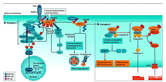
Tumor necrosis factor receptor 1 (TNFR1) signaling is a central pathway governing cell survival, inflammation, and programmed cell death. Upon tumor necrosis factor (TNF) binding, TNFR1 assembles membrane-bound complex I (containing TRADD, RIPK1, TRAF2, and E3 ubiquitin ligases like cIAP1/2). In complex I, RIPK1 primarily acts as a scaffold protein—its kinase activity is suppressed by phosphorylation and ubiquitination—to activate NF-κB, promoting pro-survival and inflammatory gene expression. When inhibitory signals are compromised, RIPK1 undergoes autophosphorylation at Ser166 (a key activation marker), transitions to cytoplasmic complex II, and triggers apoptosis or necroptosis, functioning as a "death switch."
2. Research Frontiers
Research on RIPK1 (Ser166) focuses on unraveling its precise regulation in TNFR1 signaling and its therapeutic potential. Key frontiers include defining how phosphorylation (e.g., Ser25, Ser320/335) and ubiquitination (K63/M1/K48-linked chains) suppress RIPK1 kinase activity, and how Ser166 autophosphorylation drives cell death programs. Recent advances use RIPK1 (Ser166) phosphorylation antibodies to monitor kinase activation kinetics, validate RIPK1 inhibitors in preclinical models, and explore abnormal RIPK1 activation in diseases like rheumatoid arthritis and amyotrophic lateral sclerosis. Additionally, understanding RIPK1’s role in pathogen-host interactions and tissue injury expands its translational relevance.
3. Research Significance
Elucidating RIPK1 (Ser166) regulation is critical for understanding cell fate decisions and developing treatments for inflammation- and cell death-related diseases. Ser166 autophosphorylation is the core switch converting RIPK1 from a survival scaffold to a death mediator—dysregulation contributes to autoimmune disorders, neurodegeneration, and tissue injury. RIPK1 (Ser166) antibodies serve as indispensable tools for mechanistic studies, drug pharmacodynamic evaluation, and disease subtyping, bridging basic research and clinical translation. Selective RIPK1 inhibitors, targeting Ser166-mediated activation, show promise in clinical trials, highlighting the pathway’s therapeutic potential.
4. Related Mechanisms, Research Methods, and Product Applications
4.1 Regulation of RIPK1 Kinase Activity
RIPK1 kinase activity is tightly controlled by phosphorylation and ubiquitination:
Phosphorylation-Mediated Inhibition: IKKβ/TBK1 phosphorylate RIPK1 Ser25 (blocking ATP binding), while TAK1/p38/MK2 phosphorylate Ser320/335—collectively preventing Ser166 autophosphorylation and kinase activation.
Ubiquitination-Dependent Regulation: K63/M1-linked ubiquitin chains enhance RIPK1’s scaffolding function; K48-linked chains target it for proteasomal degradation; M1-linked chains recruit autophagy proteins (ATG9, FIP200) for lysosomal clearance—all preventing abnormal activation.
4.2 RIPK1 Kinase Activation and Cell Death Programs
Ser166 autophosphorylation triggers two cell death pathways:
Apoptosis: When complex I inhibition is lifted, RIPK1 forms complex II with FADD/caspase-8. Insufficient c-FLIP enables caspase-8 homodimerization, activating caspase-3 to execute apoptosis.
Necroptosis: Inhibited caspase-8 allows activated RIPK1 to bind RIPK3, forming the necrosome. RIPK3 phosphorylates MLKL, which oligomerizes and disrupts the cell membrane, inducing inflammatory necroptosis.
4.3 Application Value of RIPK1 (Ser166) Phosphorylation Antibodies
RIPK1 (Ser166) antibodies are critical for research and drug development:
Mechanistic Studies: Monitor Ser166 phosphorylation kinetics under stimuli (TNF, pathogens) or genetic perturbations to elucidate upstream regulators and downstream effects.
Pharmacodynamic Evaluation: Serve as biomarkers to validate RIPK1 inhibitor efficacy in cellular/animal models, confirming target engagement.
Disease Research: Detect abnormal Ser166 phosphorylation in inflammatory lesions, tumors, or neurodegenerative tissues to link RIPK1 activation to disease progression.
Pathogen-Host Interaction Studies: Explore how pathogens modulate RIPK1 Ser166 phosphorylation to evade or exploit necroptosis.
4.4 Product Applications in Advanced Research
ANT BIO PTE. LTD.’s RIPK1 (Ser166) antibodies support diverse research scenarios:
Necroptosis Pathway Analysis: Detect Ser166 phosphorylation in TNF/z-VAD-fmk-induced necroptosis, validating pathway activation.
Inflammatory Disease Models: Study RIPK1 activation in ischemia-reperfusion injury, pancreatitis, or psoriasis to explore disease mechanisms.
Drug Screening: Evaluate necroptosis inhibitor (e.g., Nec-1) efficacy by monitoring Ser166 phosphorylation.
Infectious Disease Research: Investigate how viruses/bacteria modulate RIPK1 phosphorylation to influence infection outcomes.
5. Brand Mission
ANT BIO PTE. LTD. is dedicated to empowering global life science research and biopharmaceutical development through innovative, high-quality reagents. We strive to develop cutting-edge antibodies, proteins, kits, and tools that enable researchers to unravel the mechanisms of cell death, inflammation, and signal transduction. Our mission is to accelerate scientific discovery, facilitate the development of targeted therapeutics for inflammatory and neurodegenerative diseases, and improve human health by providing reliable, reproducible, and high-performance research solutions. With a commitment to excellence, innovation, and customer-centricity, we aim to be a trusted partner for researchers advancing the frontiers of precision medicine.
6. Related Product List
| Product Code |
Product Name |
Host |
| S0B6410 |
Phospho-RIP (Ser166) Recombinant Rabbit Monoclonal Antibody
(S-2702-58) |
Rabbit |
| S0B1435 |
Phospho-RIP (Ser166) Recombinant Rabbit Monoclonal Antibody
(S-1843-37) |
Rabbit |
Core Advantages of ANT BIO PTE. LTD.’s RIPK1 (Ser166) Antibodies
High Phosphorylation Site Specificity: Precisely recognize RIPK1/RIP3 Ser166 phosphorylation—a critical early marker of necroptosis initiation via the RIP1/RIP3/MLKL pathway.
Exceptional Stability and Consistency: Strict quality control ensures minimal batch-to-batch variation and reliable performance across WB, IF, and other platforms, supporting reproducible cell death and inflammation research.
7. AI Disclaimer
This article is AI-compiled and interpreted based on the original work. All intellectual property (e.g., images, data) of the original publication shall belong to the journal and the research team. For any infringement, please contact us promptly and we will take immediate action.
ANT BIO PTE. LTD. – Empowering Scientific Breakthroughs
At ANTBIO, we are committed to advancing life science research through high-quality, reliable reagents and comprehensive solutions. Our specialized sub-brands (Absin, Starter, UA) cover a full spectrum of research needs, from general reagents and kits to antibodies and recombinant proteins. With a focus on innovation, quality, and customer-centricity, we strive to be your trusted partner in unlocking scientific mysteries and driving medical progress. Explore our product portfolio today and elevate your research to new heights.
|