 中文
中文


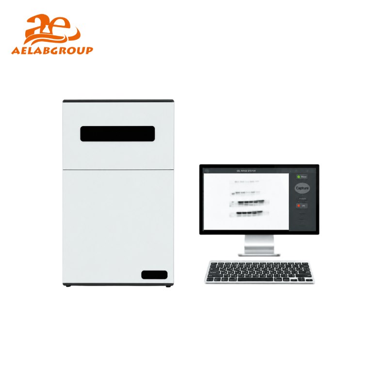
AELAB Gel and Chemiluminescence Imaging Analysis Systems

- Product Detail
- Company Profile
- DNA and RNA agarose gel imaging
- Protein gel documentation and analysis
- Western blot chemiluminescence detection
- Fluorescent nucleic acid visualization
- SDS-PAGE gel documentation
- Band intensity measurement and comparison
- Molecular weight estimation
- Gene expression and molecular biology research
- Protein expression and immunoblot studies
- Biotechnology and biopharmaceutical research
- Clinical and diagnostic research applications
- Image archiving, reporting and publication preparation
- Molecular biology laboratories
- Genetics and genomics research centers
- Protein and proteomics laboratories
- Biotechnology companies
- Biopharmaceutical research and development laboratories
- Clinical and diagnostic research laboratories
- Universities and educational institutions
- Agricultural biotechnology laboratories
- Food and environmental molecular testing laboratories
- Independent scientific research organizations
- Solutions for routine gel documentation and advanced chemiluminescence imaging
- Models for DNA, RNA, protein gel and Western blot applications
- Automatic and all-in-one imaging system options
- Digital image capture for laboratory documentation and reporting
- Systems for different sensitivity and workflow requirements
- Image analysis options for band documentation and quantitative comparison
- Technical model-selection assistance before quotation
- International supply, export packaging and shipping support
- Configuration and application support according to laboratory requirements
- Sample type: gel, membrane, DNA, RNA or protein
- Required imaging mode: fluorescence, chemiluminescence or colorimetric
- Gel or membrane dimensions
- Required detection sensitivity
- Stains, dyes or chemiluminescent substrates being used
- Required quantitative analysis functions
- Expected daily or weekly sample volume
- Preferred data export and reporting requirements
- Destination country and required power supply
AELAB provides gel documentation and chemiluminescence imaging systems for molecular biology, protein research, biotechnology, pharmaceutical research, clinical laboratories, universities and life science applications.
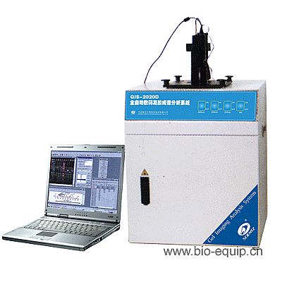
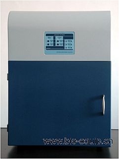
The available range includes automatic gel imaging systems, all-in-one gel documentation platforms and high-sensitivity chemiluminescence imaging systems. These instruments help laboratories visualize, capture, document and analyze DNA, RNA and protein bands after electrophoresis or membrane blotting procedures.
AELAB imaging systems are available for different research requirements, including routine gel documentation, fluorescence imaging, chemiluminescent Western blot detection and quantitative band analysis.
Compare available AELAB systems for gel documentation, fluorescence imaging, Western blot detection and molecular biology research.
Available Gel and Chemiluminescence Imaging System Types
| System Type | Typical Function |
|---|---|
| Automatic Gel Imaging System | Captures and documents stained DNA, RNA and protein gels with automated imaging and analysis functions. |
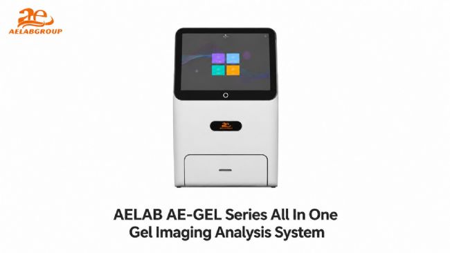
| All-in-One Gel Imaging System | Combines image capture, illumination, analysis and result documentation in an integrated laboratory platform. |
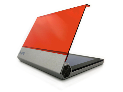
| Chemiluminescence Imaging System | Detects low-light chemiluminescent signals from Western blot membranes and related protein research applications. |
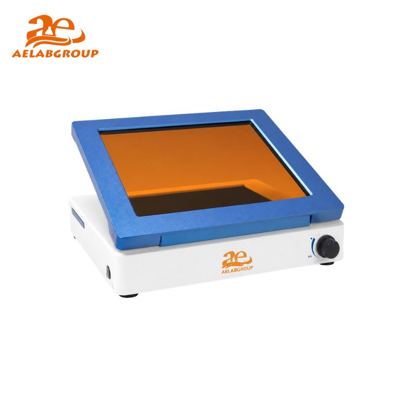
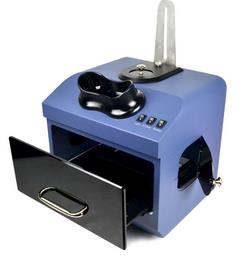
| Fluorescence Gel Imaging System | Visualizes fluorescently stained nucleic acid or protein samples using compatible excitation and detection methods. |
| Gel Documentation System | Produces digital records of electrophoresis results for comparison, reporting, publication and laboratory documentation. |
Typical Laboratory Applications
How Gel Imaging Analysis Works
1. Sample Separation
DNA, RNA or protein samples are separated through electrophoresis or transferred to a membrane.
2. Staining or Labeling
The gel or membrane is prepared with a compatible fluorescent, colorimetric or chemiluminescent detection method.
3. Image Capture
The imaging system captures the signal using the appropriate illumination source and camera configuration.
4. Data Analysis
Software is used to document bands, compare signal intensity and prepare images for reporting or publication.
How to Select the Right Imaging System
Selecting an imaging system should be based on the sample type, detection method, required sensitivity and laboratory workflow. A basic gel documentation system may be sufficient for routine nucleic acid gels, while Western blot and low-abundance protein applications may require a dedicated high-sensitivity chemiluminescence system.
| Selection Factor | Questions to Consider |
|---|---|
| Sample Type | Will the system be used for DNA gels, RNA gels, protein gels, Western blot membranes or several sample types? |
| Detection Method | Does the laboratory require fluorescence, chemiluminescence, colorimetric imaging or a combination of detection modes? |
| Sensitivity | Are routine bands being documented, or must the system detect weak and low-abundance signals? |
| Illumination Source | Which UV, blue-light, white-light or other illumination modes are required for the selected stains and assays? |
| Imaging Area | What are the dimensions of the gels, membranes or samples that must be captured? |
| Image Analysis | Are band quantification, background correction, molecular weight calculation and comparative analysis required? |
| Automation | Does the laboratory need automatic exposure, focus, image optimization or preset analysis workflows? |
| Data Export | Which image and report formats are required for archiving, publication, presentations or laboratory records? |
| Future Applications | Will the system need to support additional stains, assays or imaging modes as the laboratory develops? |
Comparison of Common Imaging Modes
| Imaging Mode | Common Samples | Typical Purpose |
|---|---|---|
| Fluorescence | DNA, RNA and fluorescently labeled proteins | Visualization and quantitative analysis of fluorescent bands |
| Chemiluminescence | Western blot membranes | Sensitive detection of enzyme-generated light signals |
| Colorimetric Imaging | Visible protein gels and stained membranes | Documentation of visible stains and colored bands |
| UV Gel Imaging | Compatible fluorescent nucleic acid gels | Routine gel visualization and documentation |
| Blue-Light Imaging | Gels prepared with compatible fluorescent dyes | Visualization with reduced dependence on UV illumination |
Who Uses AELAB Gel Imaging Systems?
Why Choose AELAB Gel Imaging Systems?
To receive a suitable model recommendation, customers should provide the following information:
AELAB is a laboratory equipment brand and supplier based in Guangzhou, China. The company supplies analytical instruments, life science research equipment, general laboratory systems and quality-control solutions to distributors, universities, research institutions and industrial customers.
The AELAB product portfolio includes gel imaging systems, electrophoresis equipment, PCR instruments, spectrophotometers, laboratory balances, environmental testing equipment, pharmaceutical testing instruments, sample preparation equipment and other laboratory solutions.
Official website: www.aelabgroup.com
Frequently Asked Questions
What is a gel imaging analysis system used for?
It is used to visualize, capture, document and analyze DNA, RNA or protein bands following electrophoresis or membrane blotting procedures.
What is the difference between a gel documentation system and a chemiluminescence imaging system?
A routine gel documentation system is commonly used to image stained electrophoresis gels. A chemiluminescence imaging system is designed to detect weak light signals, particularly from Western blot membranes and protein assays.
Can one system be used for both nucleic acid gels and Western blots?
Selected all-in-one or multi-mode systems may support several applications. The required illumination sources, filters, camera sensitivity and detection modes should be confirmed before model selection.
Which AELAB model is suitable for chemiluminescence imaging?
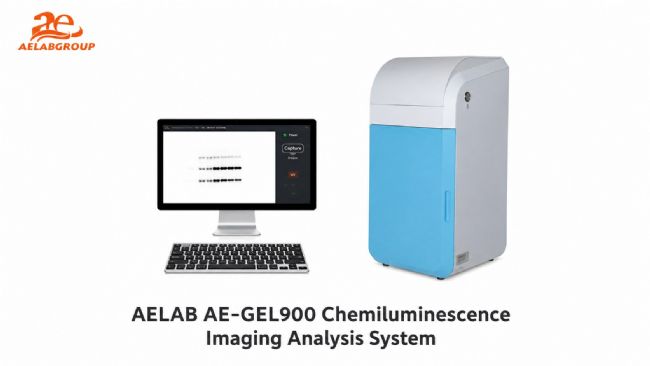
The AE-GEL900 and AE-GEL600 are included in the AELAB chemiluminescence imaging range. Final selection should be based on sample type, required sensitivity, imaging area and analysis functions.
Which models are available for routine automatic gel imaging?
The AELAB AE-GEL100 and AE-GEL200 are automatic gel imaging and analysis systems intended for efficient image capture, documentation and routine laboratory analysis.
What information is needed to select the correct imaging system?
Provide the sample type, detection method, stains or substrates, gel or membrane size, required sensitivity, analysis requirements and destination country.
Can AELAB prepare a quotation for distributors and laboratories?
Yes. AELAB can provide suitable model recommendations, technical information, available configurations, delivery details and a commercial quotation according to the inquiry.
Tell the AELAB technical sales team about your sample type, detection method, required sensitivity, imaging area, stains or substrates and analysis requirements. Suitable models can then be recommended according to your actual research workflow.
The technical proposal may include available models, specifications, optional configurations, estimated delivery time, packaging and international shipping information.
Email: info@aelabgroup.com
WhatsApp / WeChat: +86 158 0001 6110
Website: www.aelabgroup.com
-
AELAB GUANGZHOU CO., LTD.
AELAB is a laboratory equipment and analytical instrument supplier based in Guangzhou, China. We provide laboratory instruments, quality control equipment, analytical systems, sample preparation equipment, and related laboratory solutions for distributors, industrial laboratories, universities, research organizations, and testing centers worldwide.
AELAB offers both our own-brand products and carefully selected instruments from established manufacturers. OEM and ODM services are available for selected AELAB products, including customized branding, appearance, configuration, power supply, accessories, packaging, and documentation according to customer requirements.
For laboratory projects, distributor orders, government tenders, and complete laboratory setups, our team can provide a one-stop purchasing solution. We assist customers with product selection, technical comparison, quotation preparation, sourcing, quality inspection, export packaging, shipping coordination, and after-sales support.
Our technical team helps customers select suitable equipment based on their application, testing method, required specifications, budget, and local standards. Warranty and technical support are provided according to the product and project requirements.
AELAB operates under an established quality management system. Relevant company and product documentation may include SGS verification, ISO certification, CE, RoHS, FCC, or UL-related documentation, depending on the specific product model and configuration. Customers may contact us to confirm the available certificates before ordering.
Our equipment is widely used in industrial quality control, scientific research, universities, pharmaceutical laboratories, food and agricultural testing, environmental analysis, chemical laboratories, and other professional testing applications.
We are committed to developing long-term cooperation with distributors, laboratories, research institutions, and project contractors. If you are looking for a reliable laboratory equipment supplier with stable service and technical support, please contact AELAB.
Contact AELAB
Website:
https://aelabgroup.com
Email:
info@aelabgroup.com
WhatsApp / WeChat:
+86 15800016110
| Request Information |
| Other Products |
| Related Products |
| Recently viewed products |