
Reagents:
Molecular Biology
Biochemistry
Cell Biology
ELISA / Diagnostic Kits
Antibody
Serum/Medium
Other Reagents
")
- Purmorphamine (Hedgehog/Smoothened agonist)

- Product Detail
- Company Profile
- Cat.No.: SF6822-10mM
Package: 10mM×0.2ml
Description
| Cat. No. | Product Name | Package | Price (CNY) |
| SF6822-10mM | Purmorphamine (Hedgehog/Smoothened agonist) | 10mM×0.2ml | 252.00 |
| SF6822-5mg | Purmorphamine (Hedgehog/Smoothened agonist) | 5mg | 807.00 |
| SF6822-25mg | Purmorphamine (Hedgehog/Smoothened agonist) | 25mg | 2540.00 |
| Chemical Name | 9-cyclohexyl-N-(4-morpholin-4-ylphenyl)-2-naphthalen-1yloxypurin-6-amine | 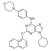 |
| Abbreviations | Purmorphamine | |
| Alias | UNII-PB12M2F8KY, Shh Signaling Antagonist VI, PB12M2F8KY | |
| Chemical Formula | C31H32N6O2 | |
| Molecular Weight | 520.62 | |
| CAS No. | 483367-10-8 | |
| Purity | 98% | |
| Solvent/Solubility | Water <1mg/ml; DMSO 4mg/ml warming; Ethanol <1mg/ml | |
| Solution Preparation | Add 0.96ml of DMSO for 5mg, or 1ml of DMSO for every 5.21mg to prepare a 10mM solution. SF6822-10mM is formulated with DMSO. |
Biological information:
| Description | Purmorphamine directly binds and activates Smoothened and blocks BODIPY-cyclopamine binding to Smo with an IC50 of approximately 1.5μM in HEK293T cells and also induces osteoblast differentiation with an EC50 of 1μM. | ||||
| Signaling Pathways | Stem Cells & Wnt; GPCR & G Protein | ||||
| Targets | Smoothened | - | - | - | - |
| IC50 | ~1.5μM | - | - | - | - |
| In vitro Studies | Purmorphamine competes with Cyclopamine (a Smo antagonist) for direct binding and activation of Smoothened, while activating the Hedgehog pathway with an IC50 of 1.5μM. Purmorphamine acts on allosteric C3H10T1/2 cells and is a potent inducer of osteogenesis. Purmorphamine acts on C3H10T1/2 cells with an EC50 (based on ALP expression) of 1 μM. Purmorphamine (1μM) in combination with BMP4 (100ng/ml) acts on 3T3-L1 cells, resulting in a more than 90-fold enhancement of ALP activity. In contrast to BMP-4, Purmorphamine acts on pluripotent mesenchymal progenitor cells and induces osteogenesis, by activating Hedgehog signaling. | ||||
| In vivo Studies | Purmorphamine acts on rat structure-based human mesenchymal stem cells to upregulate ALP expression. | ||||
| Clinical Trials | N/A | ||||
| Characterization | N/A | ||||
Relevant experimental data (this data is from published literature and Beyotime does not guarantee its validity):
| Enzyme Activity Assay Experiment | |
| Method | Smo binding experiments were performed using BODIPY-Cyclopamine and cells overexpressing Smo to obtain Smo-Myc3, deletion mutants Smo CRD (amino acid deletion at positions 68 to 182) and Smo CT (amino acid deletion at positions 556 to 793) using a CMV promoter, SV40 starting point-containing expression vector. HEK 293T cells were grown on poly-D-lysine-treated glass coverslips in 12-well plates until 70% confluence, and then transfected by appropriate expression vectors (0.5g per well) using FuGene 6. Two days after transfection, HEK 293T cells were incubated with DMEM medium containing 0.5% calf serum, 5nM BODIPY-Cyclopamine and different concentrations of Purmorphamine (0, 1.5 or 5M) (1ml per well) for 1h at 37℃. Then cells overexpressing Smo were washed using 1×PBS buffer (1 ml per well), placed in DAPI-containing medium and observed using a Leica DM4500B fluorescence microscope. For binding assays using fixed cells, HEK293T cells overexpressing Smo were fixed with 3% paraformaldehyde dissolved in 1× PBS buffer (1ml per well) for 10 min at room temperature, then treated with 1× PBS buffer (1ml per well) containing 10mM glycine and 0.2% sodium azide for 5 min and washed with 1X PBS buffer (1ml per well). The medium containing Purmorphamine was then used again for 4h at room temperature. |
| Cell Experiment | |
| Cell Line | C3H10T1/2 cells |
| Concentration | 0.5-10μM |
| Treatment Time | 4 days |
| Method | C3H10T1/2 cells were amplified in T175 flasks and passage 13 cells were isolated by trypsin/EDTA and then diluted in growth medium. Using the Multi-dropTM Liquid Transfer System, the resulting cell suspension was inoculated at 2500 cells per well into black clear bottom 384-well plates with 100µl of growth medium in the wells. After incubation overnight, cells are attached to the bottom of the wells. Using the Mini TrakTM Multi-Dispensing System, each group of Purmorphamine stock solution dissolved in DMSO (500nl) was delivered to the corresponding wells, ensuring a final concentration of Purmorphamine of 5µM. Cells were then incubated at 37℃ in air with 5% CO2. After 4 days, the medium was removed and 10μl of passive lysis buffer was added to each well. 5 min later, 10μl of alkaline phosphatase substrate solution was added to each well. After incubation at room temperature for 15 min, the experimental plates were read on an Acquest high-throughput plate reader. |
| Animal Experiment | |
| Animal Models | N/A |
| Formulation | N/A |
| Dosage | N/A |
| Administration Method | N/A |
References:
1.Sinha S, et al. Nat Chem Biol, 2006, 2(1), 29-30.
2.Wu X, et al. J Am Chem Soc, 2002, 124(49), 14520-14521.
3.Wu X, et al. Chem Biol, 2004, 11(9), 1229-1238.
4.Faghihi F, Biomed Pharmacother, 2012, 3322(12).
Packing List:
| Item | Component | Quantity |
| SF6822-10mM | Purmorphamine (Hedgehog/Smoothened Agonist) | 10mM×0.2ml |
| SF6822-5mg | Purmorphamine (Hedgehog/Smoothened Agonist) | 5mg |
| SF6822-25mg | Purmorphamine (Hedgehog/Smoothened Agonist) | 25mg |
| Manual | - | 1 copy |
Storage Conditions:
Store at -20℃, valid for at least 1 year. SF6822-5mg and SF6822-25mg can also be stored at room temperature for at least 6 months. If dissolved in non-DMSO solvents, it is recommended to store aliquots at -80℃, valid for 6 months.
Precautions:
This product is for R&D only. Not for drug, household, or other uses.
For your safety and health, please wear a lab coat and disposable gloves during the operation.
-
A leading company in China, provides kits, reagents, plastic wares and equipments for molecular biology, cell biology, biochemistry and other biomed research. Beyotime Biotech was founded in 2001 by a few Chinese scholars from Harvard and MIT, and is devoted to develop high quality kits, reagents, plastic wares and equipments for biological research.
| Request Information |
| Other Products |
| Related Products |
| Recently viewed products |