
Reagents:
Molecular Biology
Biochemistry
Cell Biology
ELISA / Diagnostic Kits
Antibody
Serum/Medium
Other Reagents

- Human VEGF ELISA Kit

- Product Detail
- Company Profile
Product Specification
| Usage | Need to bring your own test equipment 1. Microplate reader (can measure the absorption value of 450nm detection wavelength and the absorption value of 540nm or 570nm correction wavelength) 2. High-precision liquid dispenser and disposable tip 3. Distilled water or deionized water 4. Bottle washing (spray bottle), multi-channel plate washer or automatic plate washer 5. 500mL measuring cylinder 1. Preparation before the experiment 1. Sample collection and storage ① Cell culture supernatant: particulate matter should be removed by centrifugation; Test the sample immediately. If the sample is not tested in time after collection, it is recommended to pack it according to the one-time usage amount and store it in a refrigerator at-20 ℃ to avoid repeated freezing and thawing. Samples may need to be diluted with diluent (1 ×). ② Serum: Use a serum separation tube (SST) to collect samples, and place the samples at room temperature for 30 minutes. Centrifuge for 15 minutes at a rotation speed of 1000 g. The serum was removed immediately and tested immediately. If the sample is not tested in time after collection, it is recommended to pack it according to the one-time usage amount and store it in a refrigerator ≤-20 ℃ to avoid repeated freezing and thawing. Samples may need to be diluted with diluent (1 ×). ③ Plasma: Plasma was collected using EDTA, heparin or citric acid as anticoagulant, centrifuged for 15 minutes within 30 minutes after collection, at a speed of 1000g, and detected immediately. If the sample is not tested in time after collection, it is recommended to pack it according to the one-time usage amount and store it in a refrigerator ≤-20 ℃ to avoid repeated freezing and thawing. Samples may need to be diluted with diluent (1 ×). 2. Reagent preparation (Please place all reagents and samples at room temperature before use and let them stand15Minutes. All experimental samples and standards are recommendedDo repeat hole detection) ① Preparation of 1 × washing liquid: The concentrated washing liquid in the kit is 20 × mother liquid, which needs to be diluted into 1 × working liquid with distilled water before use.Example:Take 10mL of concentrated washing solution + 190mL of distilled water and make the volume to 200mL. In actual operation, the amount used can be calculated first, and then prepared. ② Preparation of 1 × dilution buffer: The concentration and dilution buffer in the kit is 10 × mother liquor, which should be diluted to 1 × working solution with distilled water before use.Example:Make up to 30 mL with 3 mL of concentration and dilution buffer + 27 mL of distilled water. In actual operation, the required amount of dilution buffer solution can be calculated according to the sample dilution factor, and then prepared. ③ Antibody detection: centrifuge the dry powder to the bottom of the tube, dissolve it with 110uL dilution buffer (1 ×), and let it stand at room temperature for 5 minutes to obtain 100 × mother liquor; Dilute to 1 × working solution before use. Calculate the required volume according to the dosage of 100uL per well.Example:After 10 wells were used, 10 uL of the detection antibody having a working concentration of 100 times was taken, and the volume was diluted to 1 mL using a dilution buffer (1 ×) to obtain 1 mL of the detection antibody having a working concentration of 1 ×. ④ SA-HRP: SA-HRP is 40 × mother liquor, which needs to be diluted with dilution buffer (1 ×) before use to prepare 1 × working solution, and the required amount per well is 100uL.Example:After 10 wells were used, 25 uL of 40 × mother liquor + 975 uL of dilution buffer (1 ×) was diluted to 1 mL to obtain 1 mL of detection antibody having a 1 × working concentration. ⑤ Developer: According to 100uL per hole, calculate the dosage required for the current test, take out the corresponding volume of developer, and protect it from light; The developer removed is for the same day use only. ⑥ Standard: The freeze-dried standard is re-dissolved with dilution buffer (1 ×), and the re-dissolving volume is 1000uL to obtain the standard mother liquor with a concentration of 2000pg/mL. Gently shake for at least 5 minutes and it dissolves well. 300 uL of dilution buffer (1 ×) was added to each dilution tube. Make serial dilutions of the standard mother liquor according to the figure below, and each tube must be thoroughly mixed before pipetting to the next tube. The standard mother solution without dilution can be used as the highest point of the standard curve (2000 pg/mL), and the dilution buffer (1 ×) can be used as the zero point of the standard curve (0 pg/mL). 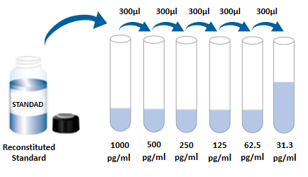 2. Operation steps 1. Prepare all required reagents and standards; 2. Take out the microplate from the sealed bag that has been balanced to room temperature. Please put the unused slats back into the aluminum foil bag and reseal them; 3. Add 300uL of washing liquid to the microplate, let it stand and soak for 30 seconds, discard the washing liquid and pat the microplate dry on absorbent paper. Please use it immediately and do not let the microplate dry; 4. Add different concentration standards, experimental samples or quality control products to the corresponding wells, 100uL per well. Sealing the reaction hole with plate sealing adhesive paper and incubating at room temperature for 2 hours; 5. Suck off the liquid in the plate and wash the plate with a bottle washer, a multi-channel plate washer or an automatic plate washer. 300 uL of washing solution was added to each well, and then the washing solution in the plate was aspirated off. Repeat the operation 3 times. Trying to absorb the residual liquid as much as possible every time you wash the plate will help to get good experimental results. At the end of the last plate washing, please suck all the liquid in the plate or turn the plate upside down, and pat all the residual liquid dry on absorbent paper; 6. Add 100 uL of detection antibody to each well. Seal the reaction wells with plate sealing tape and incubate at room temperature for 2 hours; 7. Repeat the plate washing operation in step 5; 8. Add 100uLSA-HRP to each well and incubate at room temperature for 20 minutes. Be careful to avoid light; 9. Repeat the plate washing operation in step 5; 10. Add 100uL of chromogenic solution to each microwell, incubate at room temperature for 5-30 minutes, and avoid light; 11. Add 50uL of stop solution to each microwell, and the color of the solution in the well will change from blue to yellow. If the color of the solution changes to green or the color changes are inconsistent, pat the microplate gently to mix the solution evenly; 12. Within 30 minutes after adding the stop solution, measure the absorbance value of 450nm using a microplate reader, and set 540nm or 570nm as the calibration wavelength. If dual-wavelength correction is not used, the accuracy of the results may be affected; 13. Calculation Results: Average the corrected absorbance values (OD450-OD540 or OD570), multiple well readings for each standard and sample, and then subtract the average zero standard OD value. Four-parameter logic (4-PL) curve fitting was performed using computer software to create the standard curve. Alternatively, a curve can be generated by plotting the logarithm of the standard concentration versus the logarithm of the corresponding OD value, and the best fit line can be determined by regression analysis. This process can generate a data fit that is sufficiently useful but less accurate. If the sample is diluted, the concentration should be calculated by multiplying the dilution factor.  3. Kit parameters 1. Recovery rate: Different levels of Human VEGF were spiked into cell culture medium samples and the recovery rate was determined. The recoveries ranged from 113 to 135%, with an average recovery of 121%. 2. Sensitivity: The lowest measurable dose (MDD) of Human VEGF is generally 7.8 pg/mL. The lowest measurable value is the corresponding concentration calculated from the mean of the zero-point absorbance values of 20 standard curves plus two standard deviations. 3. Calibration: This ELISA kit was calibrated with high purity recombinant Human VEGF protein expressed by sf-21 insect cells. 4. Linearity: 4 different samples were spiked with high concentrations of Human VEGF, and then the samples were diluted to the detection range with diluent (1 ×) to determine their linearity.
5. Specificity: This ELISA method can detect natural and recombinant Human VEGF protein. The following factors were configured with diluent (1 ×) at a concentration of 50 ng/mL to detect cross-reactivity with Human VEGF. Interference with Human VEGF was detected by incorporating 50 ng/mL of the interfering factor into the mid-range recombinant Human VEGF control. No significant cross-reactivity or interference was observed. The recombinant Human VEGF165/PlGF heterodimer at 3125 pg/mL showed 23% cross-reactivity and the recombinant canine VEGF at 312.5 pg/mL showed 41.2% cross-reactivity. The recombinant Human VEGF R1 (Flt-1)/Fc chimera and rhVEGF R2 (KDR)/Fc chimera interfered at concentrations > 781 pg/mL and 12.5 ng/mL. < td style = "width: 24.0699%; text-align: center; "> Recombinant human protein
4. Analysis of frequently asked questions 1. Whiteboard (after the color development is completed, no color appears)
2. Flower plate (blank and negative positive controls are normal, but the OD value of specimen wells is obviously higher)
| ||||||||||||||||||||||||||||||||||||||||||||||||||||||||||||||||||||||||||||||||
| Species Reactivity | Human | ||||||||||||||||||||||||||||||||||||||||||||||||||||||||||||||||||||||||||||||||
| Theory | This kit adopts double antibody sandwich enzyme-linked immunosorbent detection technology. Specific anti-human VEGF antibodies were pre-coated on high affinity plates. The standard substance, the sample to be tested and the biotinylated detection antibody are added to the well of the enzyme label plate, and after incubation, the VEGF present in the sample binds to the solid phase antibody and the detection antibody to form an immune complex. After washing to remove unbound material, horseradish peroxidase-labeled Streptavidin-HRP was added. After washing, a chromogenic substrate is added to protect the color from light. A stop solution was added to stop the reaction, and the absorbance value was measured at a wavelength of 450 nm (reference corrected wavelength of 540 nm or 570 nm). | ||||||||||||||||||||||||||||||||||||||||||||||||||||||||||||||||||||||||||||||||
| Synonym | MVCD1, VAS, vascular endothelial growth factor A, Vascular permeability factor, Vasculotropin, VEGF, VEGFA, VEGF-A, VEGFMGC70609, VPF, VPFvascular endothelial growth factor | ||||||||||||||||||||||||||||||||||||||||||||||||||||||||||||||||||||||||||||||||
| Composition | Please use within the expiration date of the kit
| ||||||||||||||||||||||||||||||||||||||||||||||||||||||||||||||||||||||||||||||||
| Background | Vascular endothelial growth factor (VEGF or VEGF-A), also known as vascular permeability factor, is an effective regulator of angiogenesis and angiogenesis in fetus and adults. Vascular endothelial growth factor belongs to the PDGF family, a family of proteins characterized by the presence of eight conserved cystine residues at the structure of the cystine knot and the dimer bound by antiparallel disulfide bonds. After alternative cleavage and sequence length of amino acids, humans express different isoforms, including: VEGF121, VEGF145, VEGF165, VEGF183, VEGF189 and VEGF206, among others. Among them, VEGF165 is the highest expression isoform, followed by VEGF121 and VEGF189. With the exception of VEGF121, the other isoforms contain a basic heparin-binding region and do not diffuse freely. Human VEGF165 has 88% homology to the corresponding protein amino acid sequences of mouse and rat. VEGF is expressed in various cells and tissues, including skeletal muscle cells and cardiomyocytes, hepatocytes, osteoblasts, neutrophils, macrophages, keratinocytes, brown adipocytes, CD34 + stem cells, endothelial cells, fibroblasts, vascular smooth muscle cells, and the like. The expression of VEGF is induced by hypoxia and cytokines, including IL-1, IL-6, IL-8, oncostatin M and tumor necrosis factor alpha. The expression level of VEGF is also different during development and in adults. The dimer of VEGF binds to two related tyrosine kinase receptors, namely VEGFR1 (also called Flt-1) and VEGFR2 (Flk-1/KDR). VEGF can induce both homodimerization and autophosphorylation of the latter. These receptors have seven extracellular immunoglobulin domains and one intracellular separate tyrosine receptor domain. Both vascular endothelial cells and some other non-endothelial cells express VEGF receptors. Although VEGF has the highest affinity with VEGFR1, VEGFR2 is the main factor regulating angiogenesis of VEGF. VEGF165 also binds to the semaporin receptor, Neuropilin-1, thereby promoting complex formation with VEGFR2. VEGF is known for its involvement in angiogenesis. During embryonic development, VEGF regulates the proliferation, migration and survival of endothelial cells, and thus regulates the density and volume of blood vessels. But it doesn't work on the pattern of vascularization. VEGF promotes bone formation through the recruitment of osteoblasts and chondrocytes, and it is also a monocyte chemokine. Postpartum, VEGF maintains the integrity of vascular endothelial cells and is an effective mitogen for large/small vascular endothelial cells. In adults, VEGF plays a role mainly in wound repair and the female reproductive cycle. In diseased tissues, VEGF promotes vascular permeability. Therefore, VEGF is involved in the metastatic process of tumors through extravasation and tumor angiogenesis. Various therapeutic strategies aimed at blocking VEGF activity are being used to control tumor angiogenesis induced by VEGF. The level of VEGF circulating in the body is related to the extent of autoimmune diseases (such as rheumatoid arthritis, multiple sclerosis, systemic lupus erythematosus, etc.). | ||||||||||||||||||||||||||||||||||||||||||||||||||||||||||||||||||||||||||||||||
| General Notes | 1. Please use the kit within the validity period. 2. The components of different kits and different batch kits cannot be mixed. 3. If the sample value is greater than the highest value of the standard curve, the sample should be diluted with diluent (1 ×) and re-tested; If the cell culture supernatant sample needs to be distributed and diluted, cell culture medium can be used for other intermediate dilutions except dilution with diluent in the last step. 4. Differences in test results can be caused by a variety of factors, including the operation of the experimenter, the use of the pipette, the plate washing technique, the reaction time or temperature, the storage of the kit, etc. 5. The terminating solution in the kit is an acidic solution. Please protect your glasses, hands, face and clothes when using it. 6. For scientific research only, not for in vitro diagnosis. | ||||||||||||||||||||||||||||||||||||||||||||||||||||||||||||||||||||||||||||||||
| Storage Temp. | Kit unopened, stored at 2-8 °C. | ||||||||||||||||||||||||||||||||||||||||||||||||||||||||||||||||||||||||||||||||
| Test Range | 31.3pg/mL-2000pg/mL |
-
AntBio is a biotechnology group company dedicated to serving life sciences, aiming to help scientists accelerate research and improve work efficiency. AntBio provides comprehensive and high-quality reagent tools for basic research, drug development, and diagnosis, including research grade antibodies, proteins, biochemical reagents, and assay kits. These research tools are widely used in different segments of life science research. The group company currently consists of three brands, Absin, Starter-Bio and UA-Bio.
| Request Information |
| Other Products |
| Related Products |
| Recently viewed products |