
Reagents:
Molecular Biology
Biochemistry
Cell Biology
ELISA / Diagnostic Kits
Antibody
Serum/Medium
Other Reagents

- Human Aβ ELISA Kit

- Product Detail
- Company Profile
Product Specification
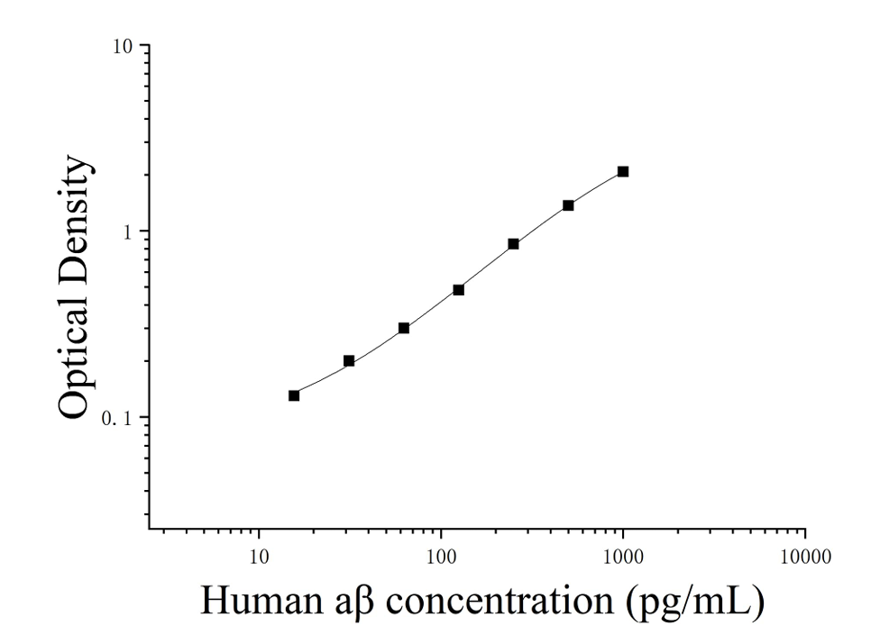
| Usage | 1. Sample processing and requirements:1. The detection range of the kit is not equal to the concentration range of the substance to be tested in the sample,It is suggested that the concentration of the test substance in the sample should be estimated by relevant literature before the experiment and the actual concentration of the sample should be determined by pre-experiment. If the concentration of the test substance in the sample is too high or too low, please dilute or concentrate the sample appropriately.2. If the sample tested is not among the sample types listed in the instructions, it is recommended to conduct pre-experiments to verify its detection effectiveness. 3. Serum: Place the whole blood sample collected in the serum separation tube at room temperature for 2 hours or 2-8 °C overnight, then centrifuge at 1000 × g for 20 minutes, take the supernatant, or place the supernatant at-20 °C or-80 °C. Store it, but avoid repeated freezing and thawing. 4. Plasma: Collect specimens with EDTA or heparin as anticoagulants, and centrifuge the specimens at 2-8 ℃ 1000 × g for 15 minutes within 30 minutes after collection. Take the supernatant for detection, or place the supernatant at-20 ℃ or-80 ℃ for storage, but avoid repeated freezing and thawing. 5. Tissue homogenate: Rinse the tissue with pre-cooled PBS (0.01 M, pH = 7.4) to remove residual blood (lysed red blood cells in the homogenate will affect the measurement results), and break the tissue into pieces after weighing. Combine the crushed tissue with the corresponding volume of PBS (generally according to the weight-to-volume ratio of 1: 9, for example, 1g of tissue sample corresponds to 9mL of PBS. The specific volume can be appropriately adjusted according to the experimental needs and recorded. It is recommended to add a protease inhibitor to PBS) into a glass homogenizer and thoroughly ground on ice. For further lysis of tissue cells, the homogenate can be sonicated, or freeze-thawed repeatedly. Finally, the homogenate was centrifuged at 5000 × g for 5-10 minutes, and the supernatant was taken for detection. 6. Cell culture supernatant: Please centrifuge at 1000 × g for 20 minutes, take the supernatant for detection, or store the supernatant at-20 °C or-80 °C, but avoid repeated freezing and thawing. 7. Cell lysate: The adherent cells were gently washed with pre-cooled PBS, then digested with trypsin, centrifuged at 1000 × g for 5 minutes and collected; The suspended cells can be collected directly by centrifugation. The collected cells were washed 3 times with pre-cooled PBS, and 150-200uL PBS was added to every 1 × 10 ^ 6 cells to resuspend (it is recommended to add protease inhibitors to PBS; if the content is very low, the PBS volume can be appropriately reduced) and the cells were disrupted by repeated freezing and thawing or ultrasound. The extract was centrifuged at 2-8 °C at 1500 × g for 10 minutes, and the supernatant was taken for detection. 8. Other biological liquids: centrifuge at 1000xg for 20 minutes, and take the supernatant for detection. 9. Sample appearance: The sample should be clear and transparent, and suspended solids should be removed by centrifugation. 10. Sample storage: If the sample is tested within 1 week after collection, it can be stored at 4 ℃. If it cannot be tested in time, please pack it according to the amount used once and store it frozen at-20 ℃ (test within 1 month), or-80 ℃ (test within 6 months) to avoid repeated freezing and thawing. Hemolysis of the sample will affect the final test results, so hemolyzed samples are not suitable for this test. 2. Preparation work before testing:1. Please remove the kit from the refrigerator 10 minutes in advance and equilibrate to room temperature.2. Preparation of standard gradient working solution: Add 1mL of universal diluent to the freeze-dried standard, let it stand for 15 minutes until it is completely dissolved, and then gently mix (the concentration is 1000pg/mL), and then dilute according to the following concentrations: 1000pg/mL, 500pg/mL, 250pg/mL, 125pg/mL, 62.5 pg/mL, 31.25 pg/mL, 15.625 pg/mL, 0pg/mL. Double dilution method: Take 7 EP tubes, add 500μL of universal diluent to each tube, draw 500μL of 1000pg/mL standard working solution into the first EP tube and mix well to prepare 500pg/mL standard working solution, according to this step, draw and mix well in sequence. The last tube is directly used as a blank hole, so there is no need to suck liquid from the penultimate tube, as shown in the figure below. 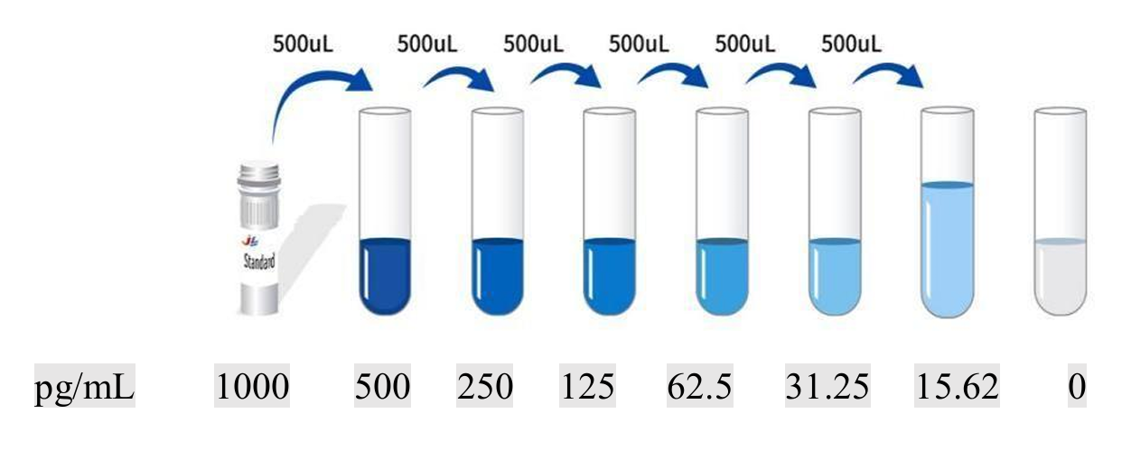 4. Preparation of enzyme conjugate working solution: Centrifuge 100 × concentrated enzyme conjugate at 1000 × g for 1 minute 15 minutes before use, and dilute 100 × concentrated HRP enzyme conjugate to 1 × working concentration with universal diluent (example: 10uL concentrated solution + 990uL universal diluent), now prepared and used. 5. Preparation of 1 × washing liquid: Take 10mL of 20 × washing liquid into 190mL distilled water (the concentrated washing liquid taken out of the refrigerator may have crystals, which is a normal phenomenon. It can be placed at room temperature and then prepared after the crystals are completely dissolved). 3. Operation steps:1. Take out the required slats from the aluminum foil bag after equilibration at room temperature for 10 minutes, and seal the remaining slats with a ziplock bag and put them back to 4 °C.2. Add samples: Add samples or standards of different concentrations to the corresponding wells at 100uL per well, and add 100uL of universal diluent to the blank wells. Incubate at 37 °C for 60 minutes after covering the plate sealing film. (Recommendation: Dilute the sample to be tested with a universal diluent at least 1 times and then add it to the enzyme labeled plate for testing. This reduces the influence of matrix effect on the test results. Finally, the sample concentration needs to be multiplied by the corresponding dilution factor when calculating. It is recommended to set up double wells for all samples and standards to be tested during testing). 3. Add biotinylated antibody: take out the enzyme labeled plate, discard the liquid, and do not wash it. 100 uL of biotinylated antibody working solution was directly added to each well, and the plate sealing membrane was covered and incubated at 37 °C for 60 minutes. 4. Plate washing: Discard the liquid, add 300uL 1x washing liquid to each well, let it stand for 1 minute, throw away the washing liquid, pat dry on absorbent paper, and repeat washing the plate 3 times (you can also use a plate washing machine to wash the plate). 5. Add enzyme conjugate working solution: add 100uL of enzyme conjugate working solution to each well, cover the sealing membrane and incubate at 37 °C for 30 minutes. 6. Wash the plate: Discard the liquid and wash the plate 5 times according to the washing method in step 4. 7. Substrate addition: 90 uL of substrate (TMB) was added to each well, covered with a plate sealing film, and incubated at 37 °C in the dark for 15 minutes. 8. Add stop solution: Take out the enzyme plate, directly add 50uL of stop solution to each well, and immediately measure the OD value of each well at a wavelength of 450nm. IV. Calculation of experimental results:Result judgment:1. Calculate the average OD value of the standard and sample replicate well and subtract the OD value of the blank well as the correction value. Taking concentration as abscissa and OD value as ordinate, the standard curve of four-parameter logic function is drawn on double logarithmic coordinate paper. 2. If the OD value of the sample is higher than the upper limit of the standard curve, it should be properly diluted and retested and multiplied by the corresponding dilution factor when calculating the sample concentration. Reference curve: The following curve is for reference only, and the experimenter needs to establish a standard curve according to his own experiment.
1. Repeatability: The coefficient of variation within the plate is less than 10%, and the coefficient of variation between the plates is less than 10%. |
 5. Kit performance
| Sample Type | Range (%) | Average recovery (%) |
| Serum (n = 8) | 84-101 | 96 |
| Plasma (n = 8) | 92-105 | 102 |
| Cell culture supernatant (n = 8) | 96-108 | 105 |
| Dilution ratio | Recovery (%) | Serum | Plasma | Cell culture supernatant |
| 1:2 | scope | 84-96 | 88-96 | 90-110 |
| Average recovery | 91 | 93 | 96 | |
| 1:4 | scope | 89-103 | 87-108 | 105-115 |
| Average recovery | 94 | 98 | 108 |
VI. Problem analysis:
If the experimental results are not good, please take photos of the color development results in time, save the experimental data, keep the used slats and unused reagents, and then contact our company's technical support to solve the problem for you. At the same time, you can also refer to the following information:| Problem Description | Possible cause | Corresponding countermeasures |
| Poor scaling curve | Incorrect dilution of standard | Ensure that the standard is dissolved and diluted according to the recommended method |
| Inaccurate pipetting | Calibrate the pipette periodically and check tip tightness | |
| Evaporation of the reaction solution | Enzyme-labeled plate is sealed with a sealing membrane | |
| Incomplete plate washing | Sufficient number of washes and addition of sufficient amount of washing liquid | |
| Foreign matter at the bottom of the hole | Clean bottom of plate before reading | |
| Weak or colorless chromogenic | Incubation time is not enough | Ensure incubation time |
| Incorrect incubation temperature | Incubate at recommended temperature | |
| Insufficient reagent volume addition | Check the pipette and follow the operation steps strictly | |
| Incorrect dilution | Test Reagent Dilution Step | |
| Enzyme conjugate inactivation | Mixed enzyme conjugate and substrate, checked by color reaction | |
| Low OD value | Incorrect plate reader settings | Check instrument wavelength |
| No stop solution added | Add an appropriate amount of stop solution | |
| Wait time too long when reading the board | Timely plate reading | |
| Excessive sample content | The appropriate dilution factor was determined by pre-experiment | |
| Sample content is too low | The appropriate dilution factor was determined by pre-experiment | |
| Background height | Contamination of chromogenic solution | Change the color developing solution |
| Color development time is too long | Controlling color development time | |
| Wrong dilution of detection antibody or enzyme conjugate | Use recommended dilution method | |
| Incomplete plate washing | Sufficient number of washes and addition of sufficient amount of washing liquid |
| Name | 9 6T match set | Storage conditions after opening |
| Pre-coated 96-well plate | 8 holes × 12 strips | -20℃ |
| Standard | 2 sticks | -20 ℃, use on the day after reconstitution |
Universal diluent | 2 × 20 mL | 2-8℃ |
Concentrated biotinylated detection antibody (100 ×) | 120uL | -20℃ |
Concentrated enzyme conjugate (100 ×) | 120uL | -20 ℃ (protected from light) |
20 × washing solution | 2 × 10 mL | 2-8 ℃ (protected from light) |
Substrate (TMB) | 10mL | 2-8℃ |
Stop liquid | 6mL | 2-8℃ |
Sealing film | 4 sheets | without |
Instructions | 1 serving | without |
2. Incorrect plate washing may lead to inaccurate results. Make sure to drain the liquid from the wells as much as possible before adding the substrate. Do not allow the wells to dry out during incubation.
3. Eliminate the residual liquid and fingerprints at the bottom of the plate, otherwise it will affect the OD value.
4. The substrate color development solution should be colorless or very light in color, and the substrate solution that has turned blue cannot be used.
5. Avoid cross-contamination of reagents and specimens to avoid wrong results.
6. Avoid direct exposure to bright light during storage and incubation.
7. Any reagent cannot come into contact with the bleaching solvent or the strong gas emitted by the bleaching solvent. Any bleaching component will destroy the biological activity of the reaction reagents in the kit.
8. Expired products cannot be used, and components with different item numbers and batch numbers must not be mixed.
9. Recombinant proteins derived from sources other than the kit may not match the antibodies in this kit and may not be recognized.
10. If it is possible to spread diseases, all samples should be managed well, and the samples and testing devices should be handled according to prescribed procedures.
-
AntBio is a biotechnology group company dedicated to serving life sciences, aiming to help scientists accelerate research and improve work efficiency. AntBio provides comprehensive and high-quality reagent tools for basic research, drug development, and diagnosis, including research grade antibodies, proteins, biochemical reagents, and assay kits. These research tools are widely used in different segments of life science research. The group company currently consists of three brands, Absin, Starter-Bio and UA-Bio.
| Request Information |
| Other Products |
| Related Products |