

- Human IL-35 ELISA Kit

- Product Detail
- Company Profile
Product Specification
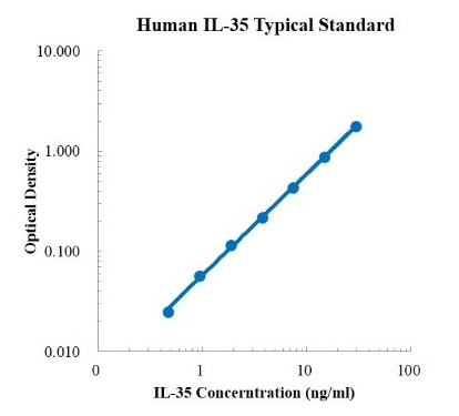
| Description | Detection Principle: This kit uses double antibody sandwich ELISA technology. The specific anti-human IL-35 capture antibody was pre coated on a high affinity microplate. Add the standard, the sample to be tested and the biotin labeled detection antibody into the wells of the enzyme plate in turn, shake well and mix well, and then place it at room temperature for 2 hours of incubation process. The IL-35 in the sample is combined with the solid-phase antibody and the detection antibody. After washing sufficiently to remove free and unbound components, streptavidin HRP (sa-hrp) labeled with horseradish peroxidase was added. After washing again, TMB chromogenic substrate was added and incubated at room temperature in the dark to develop color. The depth of color response is positively correlated with the concentration of IL-35 in the sample. Add stop solution to stop the reaction, and use a microplate reader to measure the absorbance value at 450 nm detection wavelength (correction wavelength 570-630 nm). Detection Type: Double antibody sandwich method Form: pre coated 96 well plate Test Sample Type: cell supernatant, serum, plasma Loading Amount: 100 μ L Kit Components: A copy of pre coated 96 well plate, standard, IL-35 detection antibody, standard dilution, detection buffer, TMB chromogenic substrate, washing solution, termination solution, sa-hrp, plate sealing membrane and instructions. Sensitivity: 76.93 pg/ml Detection Range: 0.47-30 ng/ml Recovery Range: 95-119% Storage Method: 2-8 ℃ Standard Curve:
Background: Interleukin 35 (IL-35)Cytokines produced by regulatory rather than effector T cells, a member of the IL-12 family, play a role in immunosuppression. It is composed of il-12α Chain and il-27β A dimeric protein composed of chains, encoded by two independent genes, il-12a and ebi3, respectively. IL-35 secreted by regulatory T cells (Tregs) inhibits the inflammatory response of immune cells. IL-35 is not continuously expressed in tissues, but after the activation of inflammatory stimuli, the gene encoding IL-35 is transcribed by vascular endothelial cells, smooth muscle cells and monocytes. Studies in mice have shown that the deletion of IL-35 chain reduces the ability of cells to suppress inflammation, which has also been observed in cell culture experiments and experimental models of inflammatory bowel disease. In order to produce inhibitory effect, IL-35 has selective activity on different T cell subsets. It can induce the proliferation of Treg cell population and reduce the activity of Th17 cell population. |
-
AntBio is a biotechnology group company dedicated to serving life sciences, aiming to help scientists accelerate research and improve work efficiency. AntBio provides comprehensive and high-quality reagent tools for basic research, drug development, and diagnosis, including research grade antibodies, proteins, biochemical reagents, and assay kits. These research tools are widely used in different segments of life science research. The group company currently consists of three brands, Absin, Starter-Bio and UA-Bio.
| Request Information |
| Other Products |
| Related Products |
| Recently viewed products |