Experimental Animal Laser Speckle Blood Flow Imager
The system is built around the emerging Laser Speckle Contrast Imaging (LSCI) technique. By virtue of its unique, non-contact, high-resolution, full-field and ultra-fast imaging capabilities, it provides clinicians and life-science researchers with a completely new and effective tool for real-time blood-flow monitoring and perfusion mapping.
Principle
LSCI extracts the velocity of moving particles (e.g., red blood cells) from the dynamic speckle pattern created when coherent laser light scatters from optically rough tissue. A CCD or CMOS camera continuously records the illuminated area, and spatial–temporal contrast analysis of the raw speckle images yields a two-dimensional map of relative blood flow without the need for any contrast agent.
Shanghai Yuyan LS-series Laser Speckle Perfusion Imagers deliver millisecond-level temporal resolution and micrometre-level spatial resolution, enabling researchers and clinicians to observe microvascular flow distribution and relative flow changes in real time. The large field-of-view optics cover everything from small animals (mice, rats) through large animals (pig, canine) to human subjects.
System Components
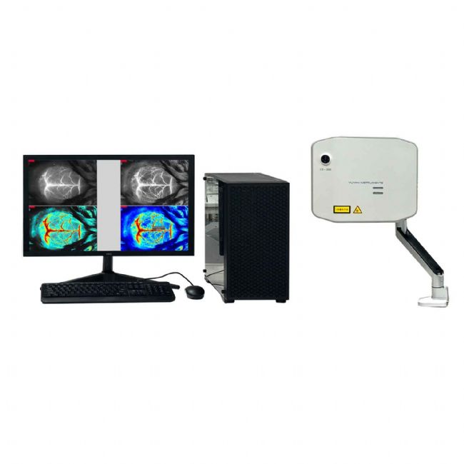
Model: LS-200 Key Features
-
Ultra-high imaging fidelity: resolves cerebral end-capillaries in mice under standard settings.
-
Industry-leading response: <200 ms latency, the fastest among comparable systems; instantaneous display of occlusion/reperfusion events.
-
Compact data footprint: ≈3 MB per file; hours of continuous recording possible thanks to 20-year algorithm optimisation.
-
One-piece microscope integration: real-time perfusion image appears while you operate under the microscope—no additional optics required.
-
Stable working distance: once focused, the field of view remains spatially locked, simplifying surgical manipulation.
-
Glare-free illumination: proprietary side-fired laser delivery eliminates specular reflection; no saline overlay needed.
-
User-friendly software: <30 min learning curve; fully optimised internal parameters; free-hand ROI selection, copy, drag, edit; automatic report generation (histograms, line graphs, tables).
Technical Highlights
Laser speckle: When coherent laser light strikes a tissue surface that is rough on the scale of the wavelength, multiply-scattered waves interfere to produce a random granular pattern—the speckle. If the scattering medium (blood cells) moves, the speckle intensity at each pixel fluctuates in time. Spatial–temporal statistics of these fluctuations are converted into quantitative flow maps.
Core Specifications
| Parameter |
LS-200 |
LS-150 |
| Laser wavelength |
785 nm |
785 nm |
| Working distance |
100 – 500 mm |
100 – 250 mm |
| Camera resolution |
2048 × 2048 |
2048 × 2048 |
| Flow-imaging speed |
>100 fps |
>100 fps |
| Focus |
Auto |
Manual |
| Imaging modes |
High-resolution & Fast |
High-resolution & Fast |
| Image registration |
Tissue outline / Colour photo / Flow overlay |
| ROI mean-flow analysis |
Select, copy, delete, drag, edit |
| TOI analysis |
Mean flow & relative change |
| Pixel density |
8.4 million pixels/cm² |
| Event marking |
Real-time flag insertion |
| Data formats |
Raw flow, standard images, video |
bio-equip.cn Contact Us:
上海玉研科学仪器有限公司
Shanghai Yuyan Instruments Co., Itd
Product Inquiries
Tel:+86-021-35183767 ,+86- 18502129044
QQ:2881513766 Wechat:Yuyanbio666
Email:sales@yuyanbio.com
Technical Support
Tel:+86-021-34173826
Email:support@yuyanbio.com
Shanghai Yuyan Instruments Co.,Ltd.is a company specializing in providing scientific instruments and technical services for animal experiments and animal research. We focus on introducing advanced scientific instruments and practical experience from abroad, and choose experimental instruments and experimental methods that are widely used, mature in application, and advanced in performance to provide scientific and reasonable solutions for the construction of domestic laboratories and the progress of research topics.
Yuyan Instruments focuses on physiology, pharmacology, toxicology and other related fields of animal experiments and research, providing customers with scientific instrument products and related services in various aspects such as animal testing, physiological functions, respiratory blood pressure anesthesia, behavior, pulmonary function research, etc. We have established good cooperation channels with many well-known scientific instrument research and development institutions in the world, and have obtained the agency rights of many famous manufacturers in China, such as: Vitalab, Pinnacle, Harvard, Starr Life, IITC, Stoelting, Union Biometrica, Stoelting and many more.
Yuyan Instruments is committed to sincerely bringing value to customers.
Our tenet: Customer's affirmation is our greatest success.
Our service philosophy: development, innovation, pragmatism, service, and provide customers with professional and high-quality services.
Our corporate mission: to provide technical products and professional services in the field of animal experiment basic research, and to make greater contributions to the scientific and technological progress and health development of the motherland.
|
