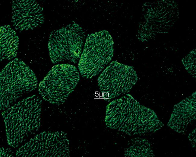
SUPER RESOLUTION
Resolve confocal images down to 120 nm XY resolution using the confocal technique and Olympus super resolution (OSR).
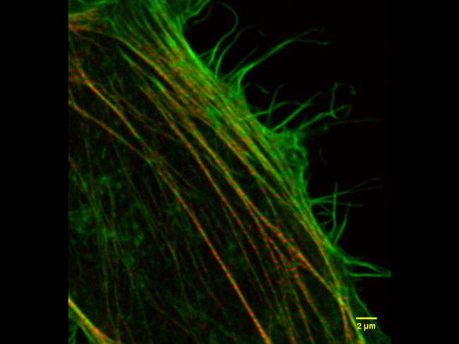
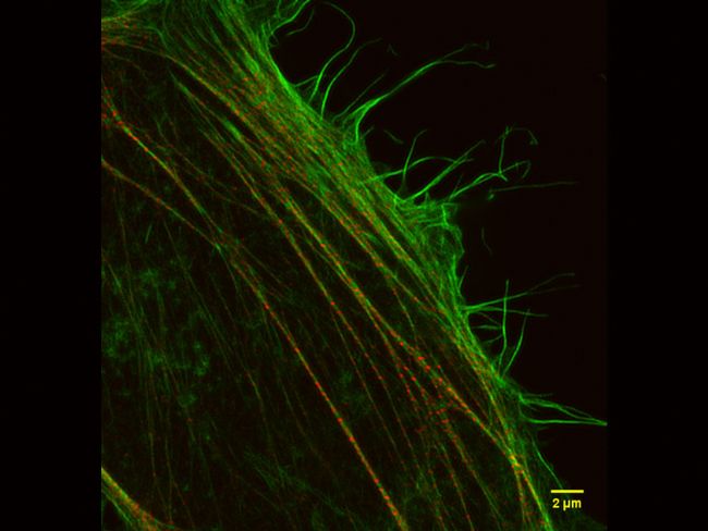
*Image: Stress fibers of Hela cell: Antibody staining with Phalloidin-Alexa488 (green) for actin, Alexa 568 (red) for myosin heavy chain. Image courtesy of: Keiju Kamijo,Ph.D. Division of Anatomy and Cell Biology, Faculty of Medicine, TOHOKU Medical and Pharmaceutical University
FAST IMAGING
Fast imaging using a spinning disk confocal and fast super resolution processing enable a live display of samples. The viability of cells during confocal time-lapse imaging is prolonged thanks to less phototoxicity and bleaching in 3D.
MULTI-MODAL
Users can easily switch between 3 modes (widefield, confocal, and super resolution).
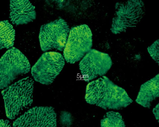
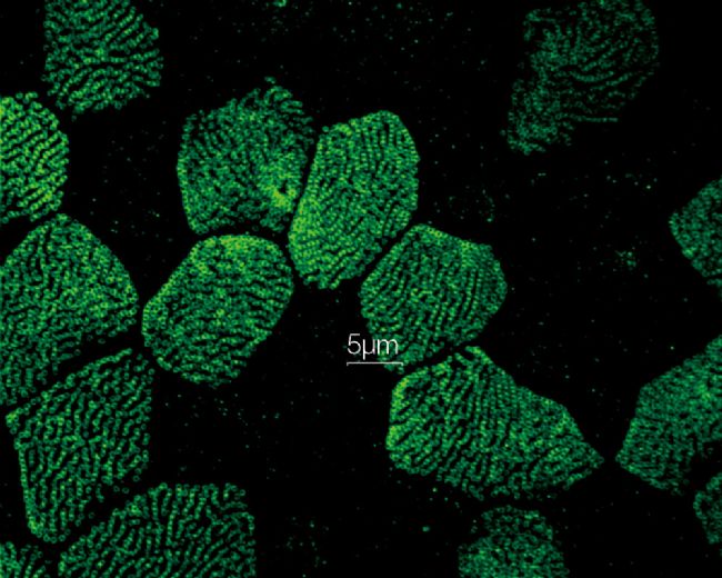
*Image: Odf2 staining (Alexa 488) of cilia at the upper part of the basal body. Image courtesy of: Hatsuho Kanoh, Elisa Herawati, Sachiko Tsukita,Ph.D. Graduate School of Frontier Biosciences and Graduate School of Medicine, Osaka University.
DEEP IMAGING

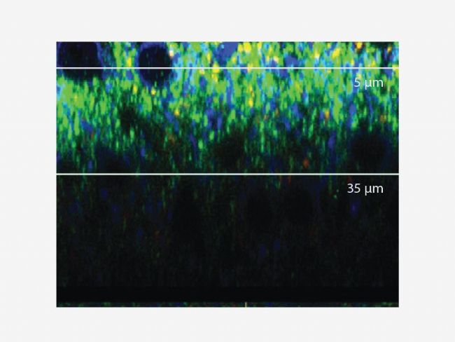
Accurate 3D reconstruction as the refraction index of silicone oil is close to that of your live sample medium.
CLEAR IMAGE
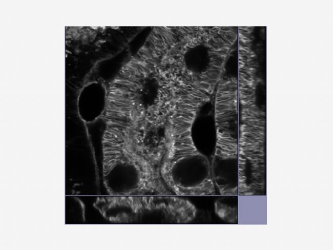
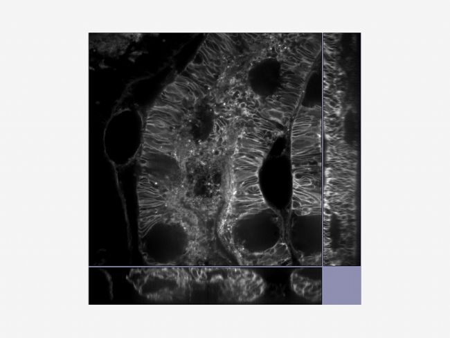
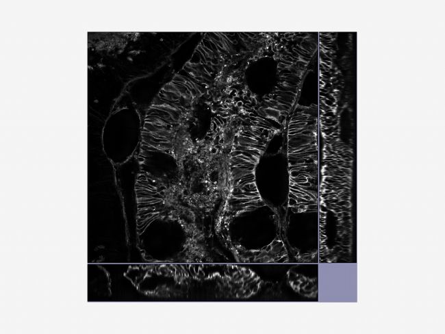
Get clear images using Olympus’ deconvolution algorithm.
*Image: Mouse kidney tissue stained with Alexa488
OSR PRINCIPLE
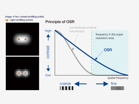
Through improved detection, specific hardware settings and signal processing, Olympus has realized improved contrast with super resolution. The Olympus SpinSR technology realizes lateral (XY) resolution down to 120 nm.
EASE OF USE
Get multi-color imaging without using specific dyes.
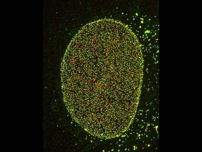
*Nuclear pore complex of Hela cell
Nup153(Alexa 488: green), Nup62(Alexa 555: red)
Image courtesy of: Hidetaka Kosako, Fujii Memorial Institute of Medical Sciences, Tokushima university
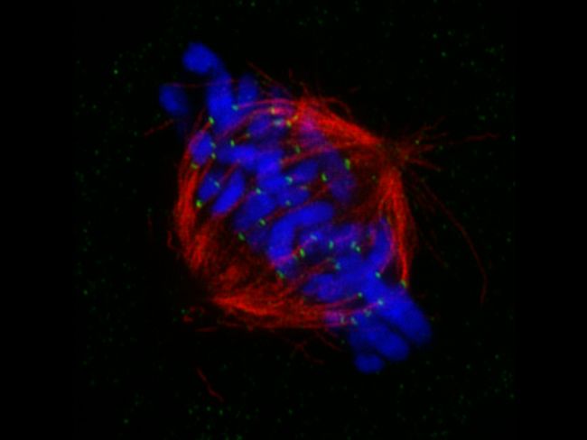
*Mitotic spindle at metapahse cell
HeLa cells derived from human cervical cancer were fixed and stained for α-tublin(microtubules,red) and Hec1(kinetochores, green),respectively. DNA was stained with DAPI(chromosomes,blue). Chromosomes interact with microtubules constituting mitotic spindle via kinetochores assembled on centromere region of chromosomes.
Image courtesy of: Masanori Ikeda and Kozo Tanaka, Department of molecular oncology,Institute of Development, Aging and Cancer
bio-equip.cn
Olympus is one of the world’s leading manufacturers of professional opto-digital products for medicine, science and industry. As a result, Olympus provides a comprehensive range of solutions. From microscopes for training and routine tasks to high-end system solutions in the fields of life science, there is a system for every need. The product line is complemented by innovative laboratory equipment for cellular research applications and the new all-in-one microscopes that offer user engagement at all levels.
