Sample Collection, Processing, and Storage Methods1. Serum: Use pyrogen- and endotoxin-free tubes. Avoid any cell stimulation during the procedure. After blood collection, centrifuge at 3000 rpm for 10 minutes to quickly and carefully separate the serum from the red blood cells. 2. Plasma: Anticoagulate with EDTA, citrate, or heparin. Centrifuge at 3000 rpm for 30 minutes and remove the supernatant. 3. Cell Supernatant: Centrifuge at 3000 rpm for 10 minutes to remove particles and aggregates. 4. Tissue Homogenate: Add an appropriate amount of saline to the tissue and mash. Centrifuge at 3000 rpm for 10 minutes and remove the supernatant. 5. Storage: If samples are not tested promptly after collection, aliquot them into single-use aliquots and freeze at -20°C to avoid repeated freeze-thaw cycles. Thaw at room temperature and ensure the sample is evenly and thoroughly thawed. Reagent Preparation: Dilution of 20× Wash Buffer: Dilute with distilled water at a ratio of 1:20, i.e., add 1 part 20× Wash Buffer to 19 parts distilled water. Plate Washing: 1. Manual Plate Washing: Shake off all liquid from the wells, fill each well with wash buffer, let stand for 1 minute, shake off all liquid from the wells, pat dry on absorbent paper, and repeat this process 5 times. 2. Automatic Plate Washer: Add 350 μL of wash buffer to each well, soak for 1 minute, and wash the plate 5 times.
Procedure 1. Remove the desired strips from the aluminum foil bag after equilibration at room temperature for 20 minutes. Seal the remaining strips in a ziplock bag and return them to 4°C. 2. Set up standard wells and sample wells. Add 50 μL of standard solution of varying concentrations to each standard well. 3. Add 10 μL of the sample to be tested to the sample wells, followed by 40 μL of sample diluent. Leave blank wells untouched. 4. Add 100 μL of horseradish peroxidase (HRP)-labeled detection antibody to each standard well and sample well, except for the blank well. Seal the wells with plate sealing film and incubate at 37°C in a water bath or incubator for 60 minutes. 5. Discard the liquid and pat dry on absorbent paper. Fill each well with wash solution, let stand for 1 minute, shake off the wash solution, and pat dry on absorbent paper. Repeat this process five times (a plate washer can also be used). 6. Add 50 μL each of substrates A and B to each well and incubate at 37°C in the dark for 15 minutes. 7. Add 50 μL of stop solution to each well. Within 15 minutes, measure the OD value of each well at a wavelength of 450 nm. Result Interpretation Draw a standard curve: In an Excel worksheet, plot the standard concentration on the horizontal axis and the corresponding OD value on the vertical axis. Draw a linear regression curve for the standard. Calculate the concentration of each sample using the curve equation. 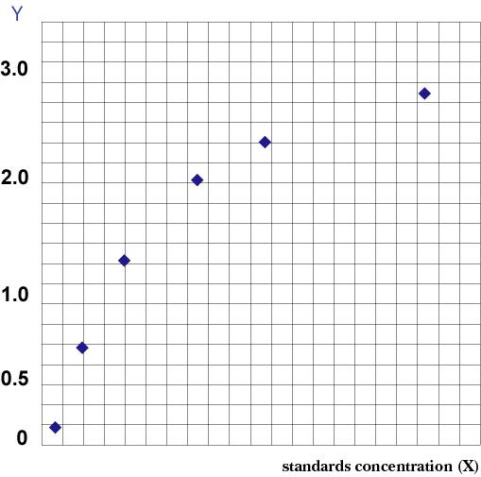 Note: Note:
- Accuracy: The correlation coefficient R value between the linear regression of the standard and the expected concentration is greater than or equal to 9900.
- Specificity: No cross-reaction with other soluble structural analogs.
- Repeatability: Both the intra-plate and inter-plate coefficients of variation are less than 15%.
| 