

- Sepax Antibodix Columns

- Product Detail
- Company Profile
- Highly cross-linked non-porous PS/DVB particle as the support
- Uniform particle size
- Particle size availability: 1.7, 3, 5, and 10 µm
- Weak-cation exchange chemistry
- Hydrophilic surface specially designed for elimination of non-specific bindings
- High separation efficiency, selectivity and resolving power
- High lot-to-lot reproducibility (link to below header)
- High loading capacity (link to below header)
- High recovery
- High mechanical stability with pressure tolerance higher than 8,000 psi for 5 and 10 µm, and 10,000 psi for 1.7 and 3 µm
- High pH stability: 2-12
- Well suited for UPLC and regular HPLC systems
- Complete selection for analytical, semi-preparative and preparative separations
- Applications for proteins, especially antibodies
Description
Antibodix columns are specially designed for high resolution, high efficiency and high recovery separations of antibodies. The packing support is composed of a rigid, spherical, highly cross-linked poly(styrene divinylbenzene) (PS/DVB) non-porous bead. The non-porous resin has particle size of 1.7, 3, 5 and 10 µm. The PS/DVB resin surface is grafted with a highly hydrophilic, neutral polymer thin layer with the thickness in the range of nanometer. On the top of the hydrophilic layer, weak cation-exchange functional groups are attached via a proprietary chemistry, resulting in high capacity ion-exchange layer.
Packing
Support:
High cross-linked, spherical PS/DVB beads
Particle size:
1.7, 3, 5 and 10 µm
Pore size:
non-porous
Phase structure:
weak cation exchanger with carboxylate functional groups chemically bonded and hydrophilic
Characteristic
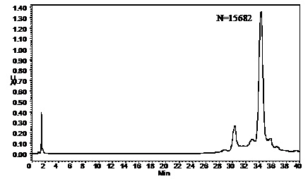
Chromatogram of a protein mixture separated by Antibodix NP5, 7.8×50mm
 |
|
Separation of MAb on Antibodix NP1.7 (1.7 µm, 4.6×50 mm)
|
 |
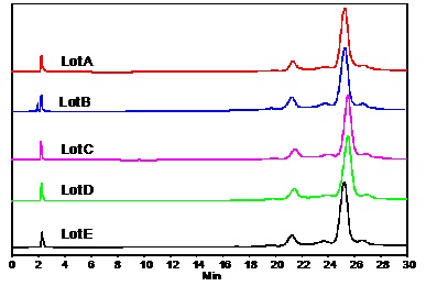
High lot to lot reproducibility
Sample 1. Protein mixtures
 |
|
Sample 2. Monoclonal antibodies
|
 |
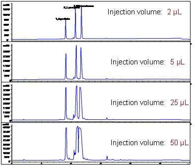
High Loading
 |
|
-
Profile
Sepax Technologies, Inc., a privately held company, was founded in Delaware, USA in November 2002. It develops and manufactures HPLC consumables, bulk media, and equipment in liquid chromatography for chemical and biological separations. It is a fast growing technology company and owns patents, proprietary technologies and know-how. Sepax has emerged as a leader in the biological separation industry in the global market.
Business strategies
Sepax Technologies, Inc. develops innovative HPLC consumables, bulk media, and instruments to solve separation challenges in the global market. We provide solution-based products by closely working with our customer scientists in pharmaceuticals, biopharmaceuticals, research institutes and government labs. Our strong technical team develops various methods with our customers to analyze complex biological molecules. As a leader in the biological separation industry, we constantly develop the best bio-separation technologies and products for our global customers. We have reached a competitive position globally in fast growing regions.
Vision Statement
To become a technology leader in the biological separation industry in the global market and provide solution based products and services for our customers.
Operation
Sepax Technologies, Inc.
Our company headquarter in Delaware is located in Delaware Technology Park with facilities of 15,000 ft2 dedicated to the development of separation resin technologies and instrumentation, production of HPLC resins, columns and CE consumables. It is also the marketing and sales center for our US and global markets.
Sales
Domestic
Audrey Fisher
Field Sales Account Manager
(Northern CA areas, UT, CO, OR)
Direct Phone: 925-324-5223
Email: afisher@sepax-tech.com
Kathleen Falls
West Coast Regional Manager
(All other CA areas, WA, TX, AZ, NM, MI)
Direct Phone: 323-228-0004
Email: kfalls@sepax-tech.com
Michael Hunnewell
Field Sales Account Manager
(MA, RI, NH, CT, NY)
Direct Phone: 302-650-3955
Email: mhunnewell@sepax-tech.com
Colleen Callahan
Midwest Account Manager
(WI, IL, IN, OH, MO)
Direct Phone: 302-366-1101, ext 106
Cell Phone: 302-339-8747
Email: ccallahan@sepax-tech.com
All other areas, please contact
Helen Gu
Vice President, Sales
Direct phone: 302-650-3909
Email: hgu@sepax-tech.com
International
Tingting Wu
Executive Account Manager/Marketing
Phone: 302-366-1101, ext 115
Email: twu@sepax-tech.com
| Request Information |
| Other Products |
| Related Products |
| Recently viewed products |