

- IAA ELISA Kit

- Product Detail
- Company Profile
Product Specification
| Usage | I. Sample Collection, Preparation, and Storage 1. Serum: After placing whole blood samples at room temperature for 2 hours or at 4°C overnight, centrifuge at 1000×g for 20 minutes. Remove the supernatant for testing. Blood collection tubes should be disposable, pyrogen-free, and endotoxin-free. Store at -20°C or -80°C and avoid repeated freezing and thawing. 2. Plasma: Within 30 minutes of collection, centrifuge at 1000×g for 15 minutes at 2-8°C. Remove the supernatant for testing. EDTA-Na2 is recommended as an anticoagulant. Avoid using samples with hemolysis or hyperlipidemia. Store at -20°C or -80°C and avoid repeated freezing and thawing. 3. Tissue Homogenization: Take an appropriate amount of tissue and wash it in pre-chilled PBS (0.01M, pH 7.0-7.2) to remove blood (lysed red blood cells in the homogenate will affect the measurement results). After weighing, mince the tissue and mix it with the appropriate volume of PBS (generally a 1:9 weight-to-volume ratio; the specific volume can be adjusted according to experimental needs and recorded. It is recommended to add protease inhibitors to the PBS). Pour the mixture into a glass homogenizer and grind thoroughly on ice. To further lyse tissue cells, the homogenate can be sonicated or freeze-thawed repeatedly (keep the sonication in an ice bath and repeat the freeze-thaw cycle twice). Finally, centrifuge the homogenate at 5000×g for 5-10 minutes. Remove the supernatant for analysis. 4. Cell Culture Supernatant: Centrifuge the cell supernatant at 1000×g for 20 minutes to remove impurities and cell debris. Remove the supernatant for testing and store at -20°C or -80°C, but avoid repeated freezing and thawing. 5. Urine: Collect the first morning urine (midstream) or 24-hour urine, centrifuge at 2000×g for 15 minutes, collect the supernatant, and store the sample at -20°C. Avoid repeated freezing and thawing. 6. Saliva: Collect the sample using a saliva sample collection tube, then centrifuge at 1000×g for 15 minutes at 2-8°C. Remove the supernatant for testing, or aliquot and store at -20°C. Avoid repeated freezing and thawing. 7. Other biological samples: Centrifuge at 1000×g for 20 minutes, remove the supernatant, and store at -20°C. Avoid repeated freezing and thawing.
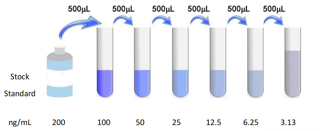
Notes: 1. The sample should be clear and transparent, and suspended matter should be removed by centrifugation. Hemolysis of the sample will affect the results, so hemolyzed samples should not be used. 2. If the sample is to be tested within one week of collection, it can be stored at 4°C. If testing cannot be performed promptly, aliquot the sample into single-use portions and freeze at -20°C (for testing within one month) or -80°C (for testing within three to six months). Avoid repeated freeze-thaw cycles. Bring the sample to room temperature before the experiment. 3. If the concentration of the test substance in your sample is higher than the highest value of the standard, please dilute it appropriately based on the actual situation (it is recommended to conduct a pilot experiment to determine the dilution factor). II.Preparation for the Test 1. Remove the test kit from the refrigerator 30 minutes in advance and equilibrate to room temperature. 2. Dilute 25 μg of concentrated wash buffer to 1 μg of working buffer with double-distilled water. Return any unused solution to 4°C. 3. Standards: Add 1.0 mL of Universal Standard & Sample Diluent to the lyophilized standard. Tighten the cap and let stand for 10 minutes until fully dissolved. Then gently mix (concentration: 200 ng/mL). Subsequently, serially dilute the standard to 200 ng/mL, 100 ng/mL, 50 ng/mL, 25 ng/mL, 12.5 ng/mL, 6.25 ng/mL, and 3.13 ng/mL. Use the standard diluent (0 ng/mL) as a blank well. Prepare the required amount of standard for use. It is recommended to add the prepared standard within 15 minutes. Do not allow it to sit for extended periods. 4. Biotin Conjugate Working Solution (1x): Centrifuge before opening the bottle. Dilute with Biotin Conjugate Diluent immediately before use. Prepare the pre-calculated total volume for each experiment (50 μL per well). Add 0.1-0.2 mL more than the actual volume. Prepare a 10 μL biotin conjugate solution with 990 μL biotin conjugate diluent, mix gently, and prepare within one hour before use. 5. Streptomycin-Horseradish Peroxidase Conjugate Working Solution (1x): Centrifuge before opening the bottle. Dilute with enzyme conjugate diluent immediately before use. Prepare the solution based on the pre-calculated total volume required for each experiment (100 μL per well). Prepare an extra 0.1-0.2 mL. Prepare a 10 μL enzyme conjugate solution with 990 μL enzyme conjugate diluent, mix gently, and prepare within one hour before use. 6. TMB Substrate - Use a pipette to aspirate the required volume of solution. Do not return any remaining solution to the reagent bottle. Notes: 1. Before using the kit, ensure that all components are dissolved and mixed thoroughly. Discard any unused standard after reconstitution. 2. Concentrated biotin conjugates and concentrated enzyme conjugates are relatively small and may disperse throughout the tube during transportation. Before use, centrifuge at 1000 × g for 1 minute to allow any liquid on the tube walls or cap to settle to the bottom. Mix the solution by carefully pipetting 4-5 times before use. Prepare the standard, biotin conjugate working solution, and enzyme conjugate working solution according to the desired volume and use the corresponding diluents. Do not mix them. 3. Concentrated wash solution may crystallize after removal from the refrigerator. This is normal. Dissolve the crystals completely in a water bath or incubator before preparing the wash solution (do not heat above 40°C). The wash solution should be at room temperature when used. 4. Samples should be added quickly, and each addition should be controlled within 10 minutes. In order to ensure the accuracy of the experiment, it is recommended to use duplicate wells. When pipetting reagents, keep the addition order consistent from one well to another. This will ensure that the incubation time of all wells is the same. 5. During the washing process, the washing solution remaining in the reaction well should be patted dry on absorbent paper. Do not place the filter paper directly into the reaction well to absorb water. Before reading, be sure to remove the residual liquid and fingerprints at the bottom to avoid affecting the reading of the microplate reader. 6. The color developer TMB should be avoided from direct exposure to strong light during storage and use. After adding the substrate, pay attention to the color change in the reaction well. If the gradient is already obvious, please stop the reaction in advance to avoid the color being too dark and affecting the reading of the microplate reader. 7. The test tubes and reagents used in the experiment are disposable. Reuse is strictly prohibited, otherwise it will affect the experimental results. 8. Please wear a lab coat and latex gloves for proper protection during the experiment, especially when testing blood or other body fluid samples. Please follow the National Biological Laboratory Safety Protection Regulations. 9. Components of the kit from different batches cannot be mixed (except for washing solution and reaction stop solution). 10. The enzyme label strips in the kit are detachable plates. Please use them in batches according to experimental needs. III.Operation Steps 1. Before starting the experiment, all reagents should be equilibrated to room temperature and all reagents should be prepared in advance. When diluting reagents or samples, they must be mixed thoroughly, and try to avoid foaming during mixing. If the sample concentration is too high, dilute it with sample diluent to bring the sample within the detection range of the kit. 2. Sample addition: Set up standard wells and wells for samples to be tested. Add 50 μL of the standard or sample to be tested, being careful not to create bubbles. Pour the sample onto the bottom of the ELISA plate well, avoiding contact with the sides. Next, add 50 μL of biotin conjugate (1 x) to each well and gently shake to mix. Cover the plate or seal the plate with film and incubate at 37°C for 1 hour. 3. To ensure the validity of the experimental results, use a fresh standard solution for each experiment. 4. After the 1-hour incubation, discard the liquid in the wells, spin dry, and wash the plate three times, adding 200 μL of wash buffer (1 x) to each well, soaking for 1-2 minutes each time, and spin dry. 5. Next, add 100 μL of streptavidin-HRP (1 x) to each well and gently shake to mix. Cover the plate or seal the plate with film and incubate at 37°C for 1 hour. 6. Discard the liquid in the wells, spin dry, and wash the plate five times, adding 200 μL of wash solution (1 x) to each well, soaking for 1-2 minutes each time, and spin dry. 7. Add 90 μL of TMB colorimetric reagent to each well in sequence and develop the color at 37°C in the dark for 15-20 minutes (shorten or extend the time as appropriate based on the actual color development, but no more than 30 minutes). 8. Add 50 μL of stop solution to each well in sequence to terminate the reaction (the blue color will immediately turn yellow). The stop solution should be added in the same order as the substrate solution. To ensure accurate experimental results, add the stop solution as soon as possible after the substrate reaction time expires. 9. Measure the optical density (OD) of each well in sequence using a microplate reader at a wavelength of 450 nm. Measure within 5 minutes after adding the stop solution. 10. *Samples may require dilution. Please see the Sample Processing section.
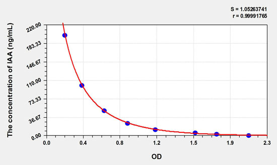
Result Calculation 1. The OD values of the competition standards and samples can be directly substituted into the calculation. If replicates are used, the average value should be used for calculation. 2. For ease of calculation, although concentration is the independent variable and OD value is the dependent variable, the graphs use the OD value of the standard as the horizontal axis (X-axis) and the concentration of the standard as the vertical axis (Y-axis). Also, to ensure intuitive visualization of the experimental results, the graphs present raw data rather than logarithmic values. Due to differences in experimental operating conditions (such as operator, pipetting technique, plate washing technique, and temperature), the OD values of the standard curve will vary. The provided standard curve is for reference only; experimenters should establish their own standard curve. The sample concentration can be calculated from the OD value of the sample used on the standard curve. This value is then multiplied by the dilution factor to determine the actual sample concentration. It is recommended to use professional curve drawing software, such as Curve Expert.
 Sample Type Recovery Range Average Recovery Serum(n=5) 83-99% 95% EDTA Plasma (n=5) 85-97% 91% heparin plasma (n=5) 85-97% Sample Type 1:2 1:4 1:8 1:16 Serum (n=5) 91-99% 83-97% 98-105% 96-101% EDTA Plasma (n=5) 85-92% 79-96% 89-101% 85-97% heparin plasma (n=5) 92-105% 86-98% 80-93% 89-97% |
Chinese name | 96T | Save conditions |
ELISA plate (removable) | 12 strips × 8 Well | 4°C/-20°C |
Lyophilized Standard | 2 | 4°C/-20°C |
Standard & Sample Dilution | 20 mL | 4°C/-20°C |
Biotin conjugate (100×) | 60 μL | 4°C/-20°C |
Biotin conjugate diluent | 10 mL | 4°C/-20°C |
Concentrated HRP Enzyme Conjugate (100×) | 120 μL | 4°C/-20°C |
Enzyme Conjugate Dilution Buffer | 12 mL | 4°C/-20°C |
Concentrated washing liquid (25×) | 20 mL | 4°C/-20°C |
Chromogenic substrate solution (TMB) | 10 mL | 4°C/-20°C (protect from light) |
Reaction stop solution | 6 mL | 4°C/-20°C |
Sealing film | 2 | Normal temperature |
1. If the entire kit is stored at -20°C, please place the kit at 4°C the night before the experiment.
2. Salt precipitation may occur when the concentrated wash solution is stored at low temperatures. When diluting, warm it in a water bath to help dissolve it.
3. A small amount of water-like substance may be present in the wells of a newly opened ELISA plate. This is normal and will not affect the experimental results.
4. This kit is for laboratory research and development use only and is not intended for use on humans or animals.
5. Reagents should be treated as hazardous substances and should be handled with care and disposed of properly.
6. Always wear gloves, lab coats, and protective glasses to avoid contact between skin and eyes with the stop solution and TMB. If contact occurs, rinse thoroughly with water.
-
AntBio is a biotechnology group company dedicated to serving life sciences, aiming to help scientists accelerate research and improve work efficiency. AntBio provides comprehensive and high-quality reagent tools for basic research, drug development, and diagnosis, including research grade antibodies, proteins, biochemical reagents, and assay kits. These research tools are widely used in different segments of life science research. The group company currently consists of three brands, Absin, Starter-Bio and UA-Bio.
| Request Information |
| Other Products |
| Related Products |
| Recently viewed products |