

- Human HA ELISA Kit

- Product Detail
- Company Profile
- Serum: Use test tubes that do not contain pyrogens and endotoxins, avoid any cell stimulation during operation. After collecting blood, centrifuge at 3000 rpm for 10 minutes to quickly and carefully separate serum and red blood cells.
- Plasma: EDTA, citrate or heparin anticoagulation. Centrifuge at 3000 rpm for 30 minutes to obtain the supernatant.
- Cell supernatant: Centrifugation at 3000 rpm for 10 minutes to remove particles and polymers.
- Tissue homogenization: Add appropriate amount of normal saline to mash the tissue. Centrifuge at 3000 rpm for 10 minutes to obtain the supernatant.
- Storage: If the sample is not tested in time after collection, please pack it according to the dosage once, freeze it at-20 ℃, avoid repeated freezing and thawing, thaw it at room temperature and ensure that the sample is evenly and fully thawed.
- Automatic plate washing machine: inject 350μL of washing solution into each well, soak for 1min, and wash the plate 5 times.
- The required slats were removed from the aluminum foil bag after equilibration at room temperature for 20 minutes, and the remaining slats were sealed with a ziplock bag and returned to 4 °C.
- First add 10μL of the sample to be tested to the sample well, and then add 40μL of the sample diluent; Blank holes are not added.
- In addition to the blank wells, 100 μL of horseradish peroxidase (HRP)-labeled detection antibody was added to each well of the standard wells and sample wells, the reaction wells were sealed with a plate-sealing membrane, and incubated in a 37 ℃ water bath or incubator for 60 minutes.
- Add 50 μL of each substrate A and B to each well, and incubate at 37 °C in the dark for 15 minutes.
- 50 μL of stop solution was added to each well, and the OD value of each well was measured at a wavelength of 450 nm within 15 minutes.
- The kit was stored at 2-8 °C and equilibrated at room temperature for 20 minutes before use. The concentrated washing liquid taken out of the refrigerator will crystallize, which is a normal phenomenon. The water bath is heated to completely dissolve the crystals before use.
- The slats not used in the experiment should be immediately put back into the ziplock bag and sealed (dry at low temperature) for storage
- Standard S0 with concentration 0 can be regarded as negative control or blank; When operating according to the instructions, the sample has been diluted 5 times, and the final result multiplied by 5 is the actual concentration of the sample.
- Carry out the incubation operation strictly according to the time, quantity and sequence indicated in the instructions.
- All liquid components are well shaken before use.
Product Specification
| Usage | Sample collection, processing and preservation methods
Preparation of reagents Dilution of 20 × wash buffer: Distilled water was diluted 1:20, i.e. 1 part of 20 × wash buffer plus 19 parts of distilled water.
Plate washing method 1. Wash the plate by hand: Remove the liquid in the hole, fill each hole with washing liquid, let it stand for 1min, then remove the liquid in the hole, pat it dry on absorbent paper, and wash the plate 5 times.
Operation steps 2. Set up standard wells and sample wells, and add 50μL of different concentrations of standards to each standard well; 5. Discard the liquid, pat dry on absorbent paper, fill each hole with washing liquid, let it stand for 1min, throw away the washing liquid, pat dry on absorbent paper, and repeat washing the plate 5 times (you can also use a plate washing machine to wash the plate).
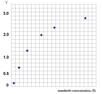
Result judgment Draw the standard curve: In the Excel worksheet, draw the linear regression curve of the standard with the concentration of the standard as the abscissa and the corresponding OD value as the ordinate, and calculate the concentration value of each sample according to the curve equation. | ||||||||||||||||||||||||||||||
| Theory | The kit uses double antibody one-step sandwich enzyme-linked immunosorbent assay (ELISA). To the coated microwells pre-coated with hemagglutinin (HA) antibody, the specimen, standard substance, and HRP-labeled detection antibody were sequentially added, incubated and thoroughly washed. The color is developed with the substrate TMB, which is converted to blue under the catalysis of peroxidase and to the final yellow under the action of acid. There was a positive correlation between the depth of color and hemagglutinin (HA) in the sample. The absorbance (OD value) was measured with a microplate reader at a wavelength of 450nm, and the sample concentration was calculated. | ||||||||||||||||||||||||||||||
| Synonym | Human hemagglutinin (HA) ELISA test kit | ||||||||||||||||||||||||||||||
| Composition |
Note: The concentrations of standard substances (S0-S5) are: 0, 50, 100, 200, 400, 800U/L in order | ||||||||||||||||||||||||||||||
| General Notes | Operational precautions
| ||||||||||||||||||||||||||||||
| Storage Temp. | Store at 2-8 ℃, shelf life is 6 months. |
-
AntBio is a biotechnology group company dedicated to serving life sciences, aiming to help scientists accelerate research and improve work efficiency. AntBio provides comprehensive and high-quality reagent tools for basic research, drug development, and diagnosis, including research grade antibodies, proteins, biochemical reagents, and assay kits. These research tools are widely used in different segments of life science research. The group company currently consists of three brands, Absin, Starter-Bio and UA-Bio.
| Request Information |
| Other Products |
| Related Products |
| Recently viewed products |