|
Breaking the Bottleneck in Glioblastoma Research: Absin Facilitates the Discovery of the Critical Link Between Plasma Cells and Cancer Stem Cells
hits:36 Date:09/02/25
Glioblastoma (GBM), as the most aggressive primary brain tumor, has long been a major challenge in the medical field. Recently, a groundbreaking study titled Infiltrating Plasma Cells Maintain Glioblastoma Stem Cells Through IgG-Tumor Binding published in the journal Cancer Cell revealed the core mechanism by which infiltrating plasma cells sustain glioblastoma stem cells (GSCs) via the IgG-FcgRIIA-AKT-mTOR axis, providing a novel target for GBM treatment.
In this high-impact research, Absin’s products served as essential tools for the research team to explore the tumor microenvironment, playing a pivotal role particularly in the immunofluorescence staining process.
Core Research Finding: The "Lethal Link" Between Plasma Cells and GBM
Using techniques such as single-cell RNA sequencing and B-cell receptor sequencing, the research team found that plasma cells (PCs) are abnormally enriched in the GBM tumor microenvironment, and this enrichment is closely associated with poor patient prognosis. These plasma cells promote tumor progression through the following mechanisms:
1. Targeted Recruitment: Recruited to the GSCs niche (stem cell niche) via the CCL2-CCR2 chemokine signal;
2. Signal Activation: The secreted IgG binds to the FcgRIIA receptor on the surface of GSCs, activating the AKT-mTOR pathway and promoting the proliferation and self-renewal of GSCs;
3. Therapeutic Potential: Blocking the stimulation of GSCs by plasma cells (e.g., targeting FcgRIIA) has become a new strategy for GBM immunotherapy.
Absin Products: The "Microscope" Illuminating the Tumor Microenvironment
In the process of analyzing the interaction between plasma cells and GSCs, immunofluorescence staining technology is a core method for visualizing cell localization and molecular expression. In the study, the research team used Absin’s Four-Color Multiplex Immunohistochemistry Staining Kit (Mouse/Rabbit Universal Secondary Antibody) (Cat. No.: abs50012), which enabled accurate detection of multiple markers in tumor tissues and provided direct morphological evidence for the key conclusions.
Analysis of Product Functions:
1. Co-Localization of Multiple Markers
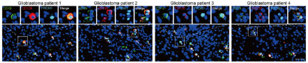
The study required simultaneous detection of plasma cell markers (CD138, PRDM1), B-cell markers (CD19), and GSC-related molecules. Absin’s four-color staining kit allowed multi-target labeling in the same tissue section, clearly demonstrating the spatial proximity between plasma cells and GSCs in the tumor microenvironment (Figure 1). This provided intuitive evidence for the conclusion that "plasma cells localize in the GSCs niche and exert their effects."
2. High Sensitivity and Specificity
Glioblastoma tissues have complex compositions, and traditional staining methods are prone to issues such as signal interference or high background. By optimizing the fluorescent labeling protocol and blocking system, Absin’s kit ensured efficient detection of low-abundance antigens (e.g., PRDM1) and accurately quantified the number of plasma cells in tumor tissues (Figure 2), laying a data foundation for the conclusion that "plasma cell enrichment is associated with poor prognosis."
3. Compatibility with Multiple Sample Types
The study involved both fresh frozen tissues and formalin-fixed paraffin-embedded (FFPE) tissues. Absin’s kit exhibited excellent compatibility with samples processed by different methods, meeting the application needs of multiple scenarios ranging from clinical samples to animal models.
Why Choose Absin?
In tumor microenvironment research, the reliability of immunofluorescence staining directly affects the credibility of conclusions. Absin’s Four-Color Multiplex Immunohistochemistry Staining Kit has become a powerful research tool due to the following advantages:
Easy Operation: The pre-prepared reagent system reduces experimental errors, allowing even novice users to master the process quickly;
Stable Signals: The fluorescent conjugates have excellent anti-bleaching performance, supporting long-term imaging analysis;
Flexible Customization: Different fluorophores can be matched according to experimental needs, adapting to various fluorescence microscopes.
This study not only reveals the pro-tumor mechanism of plasma cells in GBM but also highlights the therapeutic potential of targeting the plasma cell-GSC interaction. By enabling precise histological analysis, Absin’s products have accelerated the progress of this scientific discovery. In the future, as research on the tumor microenvironment deepens, Absin will continue to provide reliable experimental tools for fields such as immunotherapeutic target validation and drug efficacy evaluation.
Related products:
| Catalog |
Product Name |
Specification |
| abs50086 |
Absin 2-Color IHC Kit (Anti-Rabbit Secondary Antibody) |
100T |
| abs50087 |
Absin 2-Color IHC Kit (Anti-Rabbit and Mouse Secondary Antibody) |
100T |
| abs50088 |
Absin 3-Color IHC Kit (Anti-Rabbit Secondary Antibody) |
100T |
| abs50089 |
Absin 3-Color IHC Kit (Anti-Rabbit and Mouse Secondary Antibody) |
100T |
| abs50103 |
Absin 3-Color IHC Kit B (Anti-Rabbit Secondary Antibody) |
100T |
| abs50104 |
Absin 3-Color IHC Kit B (Anti-Rabbit and Mouse Secondary Antibody) |
100T |
| abs50012 |
Absin 4-Color IHC Kit (Anti-Rabbit and Mouse Secondary Antibody) |
20T/50T/100T |
| abs50028 |
Absin 4-Color IHC Kit(Anti-Rabbit Secondary Antibody) |
20T/50T/100T |
| abs50167 |
Absin 4-Color IHC Kit B (Anti-Rabbit and Mouse Secondary Antibody) |
20T/50T/100T |
| abs50168 |
Absin 4-Color IHC Kit (Anti-Rabbit Secondary Antibody) |
20T/50T/100T |
| abs50013 |
Absin 5-Color IHC Kit (Anti-Rabbit and Mouse Secondary Antibody) |
20T/50T/100T |
| abs50029 |
Absin 5-Color IHC Kit (Anti-Rabbit Secondary Antibody) |
20T/50T/100T |
| abs50014 |
Absin 6-Color IHC Kit (Anti-Rabbit and Mouse Secondary Antibody) |
20T/50T/100T |
| abs50030 |
Absin 6-Color IHC Kit (Anti-Rabbit Secondary Antibody) |
20T/50T/100T |
| abs50048 |
Absin 6-Color mlHC Kit(plus) (Anti-Rabbit Secondary Antibody) |
20T/50T/100T |
| abs50049 |
Absin 6-Color IHC Kit (plus) (Anti-Rabbit and Mouse Secondary Antibody) |
20T/50T/100T |
| abs50015 |
Absin 7-Color IHC Kit (Anti-Rabbit and Mouse Secondary Antibody) |
20T/50T/100T |
| abs50031 |
Absin 7-Color IHC Kit(Anti-Rabbit Secondary Antibody) |
20T/50T/100T |
| abs50037 |
Absin 7-Color IHC Kit (Anti-Rabbit and Mouse Secondary Antibody) |
20T/50T/100T |
| abs50038 |
Absin 7-Color IHC Kit (Anti-Rabbit Secondary Antibody) |
20T/50T/100T |
| abs50165 |
Absin 7-Color IHC Kit (Anti-Rabbit Secondary Antibody) |
20T/50T/100T |
| abs50166 |
Absin 7-Color IHC Kit (Anti-Rabbit&mouse Secondary Antibody) |
20T/50T/100T |
| abs50018 |
Absin 10-Color IHC Kit |
100T |
| abs50083 |
Lung Cancer Tumor Microenvironment mIHC Detection Kit (I) |
20T |
| abs50084 |
Lung Cancer Tumor Microenvironment mIHC Detection Kit (II) |
20T |
|