|
Guide for Inducing Colitis Organoids in Mice
hits:42 Date:09/01/25
The induction of colitis organoids in mice consists of two steps: establishment of colonic organoids and induction of colitis organoids.
Establishment of Colonic Organoids
I. Preparation Work
1. Instruments and Equipment
CO₂incubator, double-person single-sided ultra-clean bench, inverted microscope, desktop refrigerated centrifuge, water bath or water bath shaker, medical refrigerator, -80℃ refrigerator, set of pipettes, ophthalmic scissors, ophthalmic forceps.
2. Reagents and Consumables
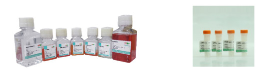
Organotial Mouse Normal Colon Organoid Culture Kit (Cat. No.: abs9985)
GFR OrganoGel Phenol red free (Cat. No.: abs9495)
60mm cell culture dish, 100μm cell strainer, 15ml centrifuge tube, several 1.5ml EP tubes, 24-well cell culture plate, metal ice box, ophthalmic scissors, ophthalmic forceps.
| Component Name |
Specification |
| Mouse normal colon organoid medium A |
100ml |
| Primary Culture Buffer B |
250ml |
| Mouse Normal Colon Primary Tissue Digestive Juice C |
30ml |
| Organoid Passage Digestive Juice D |
30ml |
| Tissue Preservation Solution E |
100ml |
| Organoid Cryopreservation Solution F |
20ml |
| Organoid Subculture Buffer G |
250ml |
II. Operation Procedure
1. Sample Preparation
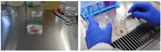
(1) Place the tissue into a sampling bottle containing pre-cooled (2-8℃) Tissue Preservation Solution E (ensure the tissue is fully submerged), and transport it back from the hospital/laboratory at 4℃.
(2) Sterilize the sampling bottle, take out the tissue, place it in a culture dish, take photos of the sample, and record information such as size, color, hardness, and tissue type.
2. Washing - Chopping
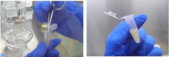
(1) Immerse the tissue in 2-3ml of Primary Culture Buffer B in a 60mm cell culture dish. Wash the tissue three times with Primary Culture Buffer B (replace the culture dish each time), then chop the tissue into pieces of approximately 1-3mm³ and transfer them to a 15ml centrifuge tube.
3. Digestion - Filtration
(2) Add 5 volumes of Primary Tissue Digestive Solution C to the 15ml centrifuge tube (volume ratio of digestive solution to tissue: 5:1; if it is difficult to estimate the tissue volume, 5mL of digestive solution is usually sufficient). Perform digestion at 4℃ for 15-30 minutes (monitor the digestion status at any time during the process).
(3) Take a small amount of the solution for observation under a microscope. When a large number of cell clusters (5-50 cells per cluster) are observed, add 3 volumes of Primary Culture Buffer B (volume ratio of buffer to digestive solution: 3:1) to terminate digestion. Gently pipette the solution with a pipette tip, and the solution will become turbid.
(4) Filter the solution using a 100μm cell strainer, and observe a small amount of the filtrate under a microscope. Collect the filtrate into a 15ml centrifuge tube, centrifuge at 300×g and 4℃ for 5 minutes for enrichment, then remove the supernatant.
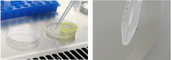
4. Matrigel Addition - Plating - Medium Addition (This is the key step of the entire primary operation)
(1) Preparation Work
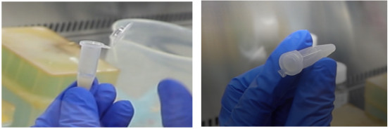
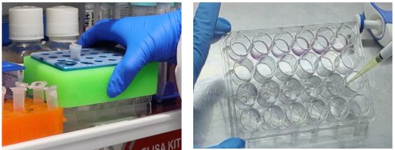
a. Matrigel should be placed in a metal ice box and thawed overnight in a 4℃ refrigerator.
b. Pipette tips and centrifuge tubes need to be pre-cooled at -20℃ for at least 30 minutes.
c. Thawed Matrigel can be stored at 4℃ and is recommended to be used within 2 weeks.
(2) Seeding Requirements
Use a 24-well plate; add 25μl of Matrigel-cell cluster mixture and 500-750μl of organoid medium to each well.
(3) Seeding Density
Density Recommendation 1: Volume ratio of Matrigel to cell cluster pellet = 25:1 (if it is difficult to estimate the volume of the cell cluster pellet, adding 300μl of Matrigel is usually sufficient).
Density Recommendation 2: 500 cell clusters per 25μl of Matrigel (this density can be used as a reference if cell counting is required for seeding).
(4) Matrigel Addition - Plating
Add Matrigel (Cat. No.: abs9495) to the cell cludster pellet, pipette gently to mix (avoid full pipetting to prevent air bubbles), then perform plating. The entire operation should be carried out on a metal ice box or ice. For skilled operators, the process of Matrigel addition, mixing, and plating should be completed within 30 seconds to maintain good fluidity of the Matrigel.
(5) Medium Addition
Place the plated culture plate in a 37℃ incubator for 40-60 minutes to allow Matrigel solidification, then add 500-750μl of Organoid Medium A for culture. After approximately 10-14 days, most organoids will reach a diameter of 200μm-500μm and can be passaged.
Induction of Colitis Organoids
I. Preparation Work
Mouse Normal Colonic Organoid Medium
Organotial Mouse Normal Colon Organoid Culture Kit (Cat. No.: abs9985)
GFR OrganoGel Phenol red free (Cat. No.: abs9495)
DSS (Dextran Sodium Sulfate)
Mouse IL-6 ELISA Kit (Cat. No.: abs520004)
Mouse TNF-α ELISA Kit (Advanced), 60mm cell culture dish, 100μm cell strainer, 15ml centrifuge tube, several 1.5ml EP tubes, 24-well cell culture plate, metal ice box.
II. Induction Method of Colitis Organoids
For passaged organoids, when organoid primordia (with vesicles) appear on Day 2-3, treat them with organoid medium containing 20μg/mL DSS for 120 hours continuously, and replace the DSS-containing organoid medium every 2 days during this period.
III. Verification of Colitis Organoids
1. Bright-Field Verification
Record bright-field photos and videos of organoids in the Control group (non-inflammation-induced group) and Model group (inflammation-induced group) every day.
Compared with the Control group, the Model group shows organoids with loose morphological structure and swelling.

Bright-field image observation: Compared with the Control group, the Model group shows organoids with significantly thickened walls.
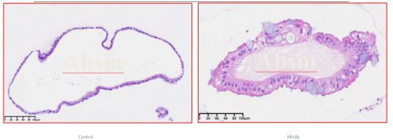
2. HE Staining
HE staining results: Compared with the Control group, the Model group shows organoids with thickened walls, disordered epithelial arrangement, and a certain degree of hyperplasia and intracellular vesicle structures.
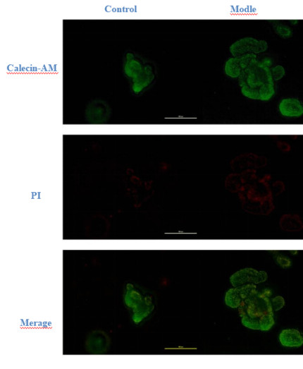
3. Organoid Live/Dead Staining
Live/dead staining results: Compared with the Control group, the Model group shows organoids with decreased viability.
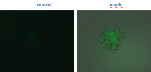
4. FITC-Dextran Fluorescence Staining
FITC-dextran fluorescence staining results: Compared with the Control group, the Model group shows organoids with increased permeability.
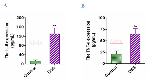
5. Detection of IL-6 and TNF-α
Detection results of IL-6 and TNF-α: Compared with the Control group, the Model group shows organoids with increased permeability; ELISA detection shows significantly elevated IL-6 levels in the Model group (Figure A) and significantly elevated TNF-α levels in the Model group (Figure B).
Conclusion: By comparing the indicators of the Control group (non-inflammation-induced group) and the Model group (inflammation-induced group) through bright-field verification, HE staining, organoid live/dead staining, FITC-dextran fluorescence staining, and detection of IL-6 and TNF-α, it can be verified that inflammation induction in the Model group is successful.
Related products:
| Product Name |
Catalog |
Specification |
| Organotial Mouse Normal Colon Organoid Culture Kit |
abs9985 |
1kit |
| GFR OrganoGel Phenol red free |
abs9495 |
1.5ml*8 |
| Mouse IL-6 ELISA Kit |
abs520004 |
96T |
|