 中文



Home > News > What is the Key Role of FOXP3 Antibody in Regulatory T Cell Research?
What is the Key Role of FOXP3 Antibody in Regulatory T Cell Research?
- FOXP3, as the key transcription factor of regulatory T cells (Treg cells), plays a decisive role in the development, differentiation, and functional maintenance of Treg cells.
1. How does FOXP3 regulate the development and function of regulatory T cells?
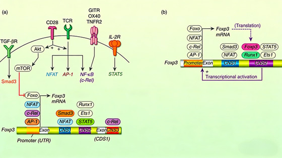
FOXP3, as the key transcription factor of regulatory T cells (Treg cells), plays a decisive role in their development, differentiation, and functional maintenance. During the precursor stage of Treg cells, the expression of FOXP3 is finely regulated by multiple signaling pathways. T cell receptor signals activate transcription factors such as NFAT, AP-1, and NF-κB, which cooperate with AML1/Runx1 to act on the promoter and conserved non-coding sequence 3 (CNS3) region of the FOXP3 gene, initiating its initial transcription. However, CD28 signaling-mediated activation of the Akt and mTOR pathways inhibits FOXP3 expression, explaining why strong co-stimulatory signals may hinder Treg differentiation. In mature Treg cells, FOXP3 maintains stable expression through a positive feedback loop. The FOXP3 protein forms a complex with AML1/Runx1 in the conserved non-coding sequence 2 (CNS2) region, inducing DNA demethylation in this region. Together with STAT5 activated by the IL-2 signaling pathway, it consolidates FOXP3 transcription, ensuring the stable phenotype and function of Treg cells.
2. What is the relationship between FOXP3 protein structure and function?
The FOXP3 protein contains multiple functional domains, and its unique structural features determine its transcriptional regulatory functions. A specific linker region, known as the Runx1-binding region, exists before the forkhead domain, enabling FOXP3 to form a head-to-head dimer configuration. This special dimerization not only endows FOXP3 with specific DNA-binding properties but also provides a binding site for the cofactor Runx1, jointly regulating the transcription of downstream target genes. Structural studies have shown that this head-to-head dimer configuration is crucial for FOXP3's transcriptional activation function and is essential for Treg cells to maintain immune homeostasis. Conversely, when FOXP3 forms a domain-swapped dimer, its transcriptional activity is significantly impaired, leading to the loss of Treg cell function. This structure-function relationship provides a molecular basis for understanding FOXP3's regulatory mechanisms.
3. What are the main applications of FOXP3 antibodies in basic research?
FOXP3 antibodies, as important tools for studying Treg cells, have broad applications in basic immunology research:
01. Cell identification and sorting: Using flow cytometry combined with FOXP3 antibodies, FOXP3+ Treg cells can be accurately identified and sorted, which is fundamental for studying their biological properties.
02. Tissue localization analysis: Immunohistochemistry with FOXP3 antibodies can reveal the distribution of Treg cells in different tissues, particularly their infiltration characteristics in tumor microenvironments, inflammatory sites, and autoimmune lesions.
03. Protein expression quantification: Techniques such as Western blot and ELISA, using FOXP3 antibodies, can quantitatively analyze FOXP3 protein expression levels and study its dynamic changes under various physiological and pathological conditions.
04. Protein interaction studies: Combined with co-immunoprecipitation and chromatin immunoprecipitation, FOXP3 antibodies can explore the interaction networks between FOXP3 and other transcription factors, co-regulators, and chromatin-modifying proteins.
4. What is the value of FOXP3 antibodies in clinical diagnosis and disease research?
In clinical medicine, FOXP3 antibodies have become important tools for studying various diseases:
01. Autoimmune disease research: Detecting the quantity and distribution of Treg cells in lesions can assess changes in immune tolerance status, providing critical insights into the pathological mechanisms of autoimmune diseases such as systemic lupus erythematosus and rheumatoid arthritis.
02. Tumor immunology research: In various solid tumors and hematological malignancies, FOXP3 antibodies can evaluate the abundance of tumor-infiltrating Treg cells, which are often associated with immunosuppressive microenvironments and poor prognosis.
03. Transplant immunology monitoring: After organ transplantation, monitoring changes in FOXP3+ Treg cells in peripheral blood or graft tissues can assess immune tolerance status, offering references for predicting and intervening in rejection reactions.
04. Diagnosis of immunodeficiency diseases: In primary immunodeficiency diseases such as IPEX syndrome, FOXP3 gene mutations lead to the absence or dysfunction of FOXP3 protein. FOXP3 antibody detection aids in the diagnosis and classification of such diseases.
5. What are the application prospects of FOXP3 antibodies in drug development?
With a deeper understanding of the role of Treg cells in diseases, FOXP3 antibodies show multifaceted potential in drug development:
01. Targeted therapy development: Functionally modulatory antibodies designed for specific disease states may achieve therapeutic goals by regulating FOXP3 activity or Treg cell function.
02. Drug screening platform construction: FOXP3 antibody-based detection systems can screen small-molecule compounds that modulate Treg cell differentiation or function, providing new drug candidates for autoimmune diseases, allergies, and tumor immunotherapy.
03. Quality control in cell therapy: In the development of Treg cell therapy products, FOXP3 antibodies are key quality control tools for assessing cell purity, stability, and functional status.
04. Immunotherapy efficacy prediction: In cancer immunotherapy, FOXP3 expression levels may serve as biomarkers for predicting the efficacy of immune checkpoint inhibitors. The development of related antibodies aids in formulating personalized treatment plans.
6. Which manufacturers provide FOXP3 antibodies?
Hangzhou Start Biotech Co., Ltd. has independently developed the "Alexa Fluor® 488 Mouse Anti-Human FOXP3 Antibody (S-2903)" (Catalog No.: S0B5822), a high-fluorescence-brightness, excellent nuclear staining performance, and highly stable antibody for flow cytometry and immunofluorescence detection. This product uses high-quality mouse monoclonal antibodies, labeled with Alexa Fluor® 488 fluorescent dye through optimized conjugation processes, and undergoes special formulation treatment. It efficiently and specifically penetrates the nuclear membrane of fixed/permeabilized cells to recognize human forkhead box protein P3 (FOXP3), making it an excellent tool for the precise identification and analysis of human regulatory T cells (Treg).
Core Product Advantages
Bright fluorescent signal and excellent photostability: Labeled with Alexa Fluor® 488 dye, it offers higher fluorescence brightness and better photostability compared to traditional FITC. It is less prone to quenching in multicolor flow analysis or prolonged imaging, providing more stable and sensitive detection signals to effectively distinguish FOXP3-positive and negative cell populations.
Efficient nuclear staining and exceptional specificity: This product is optimized with a formulation for nuclear antigen detection. Under standard fixation/permeabilization conditions, it efficiently enters the nucleus and specifically binds to the FOXP3 transcription factor. Its specificity has been rigorously validated, ensuring accurate and reliable staining results. It is the gold standard tool for identifying FOXP3+ Treg cell subsets in human CD4⁺CD25⁺ T cells.
Ready-to-use convenience and outstanding batch consistency: The product is provided in a ready-to-use buffer, pre-titrated for optimal performance, allowing direct use in intracellular staining steps after fixation/permeabilization. Strict production quality control ensures high consistency in fluorescence intensity, staining index, and specificity across batches, guaranteeing long-term comparability of experimental data.
Key Research and Clinical Applications
Precise identification and quantification of regulatory T cells (Treg): When used in combination with surface marker antibodies such as anti-CD4 and anti-CD25, it accurately quantifies the proportion of functional Treg cells in human peripheral blood, tissues, or in vitro cultured cells through flow cytometry or immunofluorescence techniques. This is critical for assessing immune homeostasis and tolerance status.
Autoimmune diseases and immune dysregulation research: Used to study changes in the quantity and function of Treg cells in patients with systemic lupus erythematosus (SLE), rheumatoid arthritis (RA), type 1 diabetes, etc., exploring the immune mechanisms underlying disease development.
Tumor immune microenvironment analysis: Used to evaluate the abundance of Treg cells in tumor-infiltrating lymphocytes (TILs) or peripheral blood. Their levels are associated with immunosuppressive microenvironments and serve as potential biomarkers for predicting the efficacy and prognosis of immunotherapies (e.g., PD-1 inhibitors).
Transplant immunology and immunosuppression research: Monitors changes in Treg cells in transplant patients or before/after immunosuppressive therapy, assessing immune status and transplant rejection risks.
Immunotherapy development and evaluation: Serves as a key indicator in drug development or cell therapies (e.g., CAR-T) to assess the impact of therapeutic strategies on Treg cells.
Related News
- What is the Key Role of FOXP3 Antibody in Regulatory T Cell Research? 11/3/2026
- Summary of Immunohistochemistry (IHC) Kits in Modern Pathological Diagnosis 11/2/2026
- Unraveling IL‑5‑Mediated Immune Regulation and Disease Pathogenesis 11/1/2026
- How Rabbit ELISA Kits Deliver High Sensitivity and Specificity for Protei 10/30/2026
- Optimizing Cell Experiment Quality with Dead Cell Removal Magnetic Beads 10/29/2026
- Dead Cell Removal Sorting: Elevating Experimental Data Quality in Cell Research 10/28/2026
- Cytokine Antibodies: Decoding Regulatory Networks in Autoimmune Diseases 10/27/2026
- Dead Cell Removal Sorting: A Key Strategy to Enhance Experimental Data Reliabili 10/26/2026
- TH Recombinant Rabbit Monoclonal Antibody: Advances in TH Deficiency Diagnosis a 10/25/2026
- Human Angiotensin-Converting Enzyme 1 (ACE1): Core Catalytic Traits, Regulatory 10/23/2026