
Home > News > Single-Cell + Spatial Transcriptomics Reveal the Key Axis of PTMC Progression
Single-Cell + Spatial Transcriptomics Reveal the Key Axis of PTMC Progression
- Recently, a landmark study on the progression mechanism of papillary thyroid microcarcinoma (PTMC) was published in Advanced Science (2025). The team from China Medical University, using multi-technical approaches such as single-cell RNA sequencing (scRNA-seq) and spatial transcriptomics, identified for the first time that the PROS1-MERTK axis is the core pathway driving the crosstalk in the PTMC microenvironment and disease progression. In the critical validation phase of protein expression and colocalization, the research team selected Absin's Multiplex Fluorescence Immunohistochemistry Kit (Catalog No.: abs50014), successfully achieving accurate visualization of multi-target proteins and providing direct evidence for mechanism elucidation.
Literature Title: PROS1‐MERTK Axis Drives Tumor Microenvironment Crosstalk and Progression in Papillary Thyroid Microcarcinoma
Published Journal: Advanced Science (IF 14.1)
DOI: https://doi.org/10.1002/advs.202413474
Core Reagent: Six-Color Multiplex Fluorescence Immunohistochemistry Staining Kit (Mouse/Rabbit Universal Secondary Antibody) (abs50014)
I. Research Background: Clinical Pain Points of PTMC Urgently Require Mechanistic Breakthroughs
The incidence of papillary thyroid carcinoma (PTC) has been increasing year by year, among which papillary thyroid microcarcinoma (PTMC, diameter ≤ 1 cm) accounts for more than 50%. Although most PTMCs grow slowly with a favorable prognosis (10-year survival rate of 93.5%-97%), 0.4%-28.8% of cases still experience rapid progression (e.g., tumor enlargement, lymph node metastasis, extrathyroidal invasion). However, clinically, there is a lack of biomarkers and intervention targets for accurately identifying progression risks.
In addition, since the concept of "PTMC overdiagnosis and overtreatment" was proposed in 2013, countries such as Japan and the United States have promoted "active surveillance (AS)" as an alternative to surgery. Nevertheless, distinguishing between "indolent PTMC" and "progressive PTMC" remains a clinical challenge—which is precisely the core objective of this study: to explore the key molecular mechanisms and potential biomarkers of PTMC progression.
II. Core Research Idea: Systematic Elucidation from "Cellular Ecology" to "Pathway Mechanism"
The research team adopted a logical chain of "multi-dimensional sequencing + multi-level validation" to progressively uncover the mechanism of PTMC progression. The specific ideas are as follows:
1. Sample Design: Covering All Stages of the Disease to Ensure Data Representativeness
Nineteen surgical specimens (from 15 patients) were selected, covering four key groups:
* Normal thyroid tissue (4 cases)
* Non-progressive PTMC (4 cases, no progression after 5 years of AS)
* Progressive PTMC (5 cases, with tumor enlargement/metastasis)
* Progressive PTC (6 cases, advanced cases)
2. Technical Route: Three-Step Process of "Sequencing Analysis → Spatial Localization → Functional Validation"
(1) Single-cell level: scRNA-seq was performed on 146,529 cells, which were divided into 32 cell clusters. The dynamic changes of 6 major cell types, including thyroid cancer cells, fibroblasts, and immune cells, were identified;
(2) Spatial level: Combined with 10x Visium spatial transcriptomics, the distribution of different cell populations in tissues was localized, and the heterogeneity of progressive tumors was analyzed;
(3) Communication level: CellChat was used to analyze intercellular ligand-receptor interactions, and the PROS1-MERTK axis was identified as the key pathway;
(4) Protein validation level: Absin's Multiplex IHC/IF Kit (abs50014) was used to detect the expression and colocalization of target proteins, verifying the cellular origin of the pathway;
(5) Functional validation level: Cell migration/invasion assays and mouse subcutaneous xenograft models were used to confirm the progression-promoting effect of the PROS1-MERTK axis;
(6) Mechanism exploration level: SCENIC analysis and dual-luciferase reporter assay were used to identify the transcription factors (NFYB/FOXP2) regulating PROS1 and downstream pathways (WNT/TGF-β).
III. Key Research Results: The PROS1-MERTK Axis Serves as an "Accelerator" for PTMC Progression
Through systematic analysis, the research team drew 4 core conclusions, each supported by solid data:
1. PTC Progression Is Accompanied by "Immune Suppression + Fibroblast Enrichment" in the Microenvironment
scRNA-seq results:
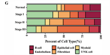
* With disease progression (normal → Stage I → Stage II → Stage III), the proportion of T/NK cells gradually decreased (enhanced immune suppression);
* The proportion of fibroblasts increased significantly (from approximately 5% in normal tissues to 18% in Stage III), becoming the core "signal senders" in the microenvironment.
2. The PROS1-MERTK Axis Is the "Key Hub" of Intercellular Communication
Analysis of intercellular ligand-receptor interactions via CellChat revealed:
* PROS1 (ligand) is mainly secreted by "adipogenic cancer-associated fibroblasts (adi-CAFs)" in the progressive stage;
* MERTK (receptor) is mainly expressed in progressive tumor cells and myeloid cells (e.g., tumor-associated macrophages);
* The interaction intensity significantly increases with progression (normal < Stage I < Stage II ≤ Stage III), and promotes progression through two modes: paracrine (CAFs → tumor cells) and autocrine (tumor cells autocrine PROS1).
3. NFYB/FOXP2 Regulate PROS1 Transcription as the "Upstream Switch" of the Pathway
SCENIC transcription factor analysis and dual-luciferase assay showed:
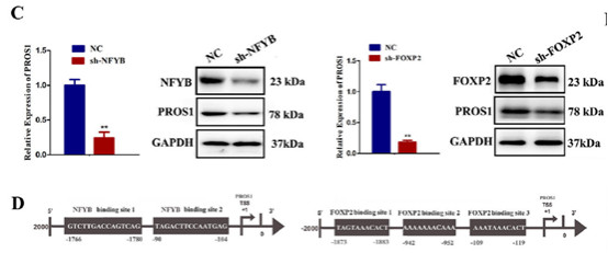
* In fibroblasts, NFYB and FOXP2 can directly bind to the PROS1 promoter and activate its transcription;
* After knocking down NFYB/FOXP2, the mRNA and protein expression of PROS1 decreased by more than 60%, and the migration ability of tumor cells was significantly weakened.
4. The PROS1-MERTK Axis Promotes Progression via the WNT/TGF-β Pathway, and MERTK Can Serve as a Biomarker
Western blot and GSEA analysis confirmed:
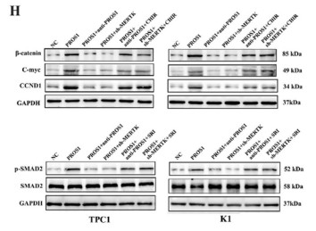
* After PROS1 binds to MERTK, it activates the downstream WNT/β-catenin and TGF-β/SMAD2 pathways, promoting tumor cell proliferation and invasion;
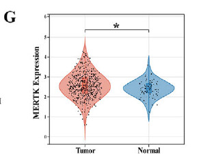
* TCGA database validation: MERTK is highly expressed in progressive PTMC/PTC, and patients with high MERTK expression have a poorer prognosis (disease-free survival shortened by 35%), suggesting that MERTK can serve as a potential biomarker and therapeutic target for PTMC progression.
IV. Support from Absin Products: Multiplex Fluorescence Kit Solves the "Protein Localization Problem"
In the entire study, "the cellular origin of PROS1" and "the tissue localization of PROS1-MERTK" were key scientific issues, and Absin's Multiplex IHC/IF Kit (abs50014) was the core tool to solve this problem.
1. Product Information and Application Scenarios
| Product Name | Catalog No. | Core Advantages | Application Scenarios in the Study |
| Six-Color Multiplex Fluorescence Immunohistochemistry Staining Kit (Mouse/Rabbit Universal Secondary Antibody) | abs50014 | 1. Multi-target compatibility (capable of detecting 3-4 proteins simultaneously); 2. High signal specificity and low background; 3. Compatible with laser confocal microscopy imaging | Detection of the expression and colocalization of proteins such as PROS1, ACTA2, and MERTK in tissue sections |
2. Specific Role in the Study: Directly Verifying That "adi-CAFs Are the Main Source of PROS1"
The research team used the abs50014 kit to perform multiplex immunofluorescence staining on tissue sections at different stages, obtaining key evidence:
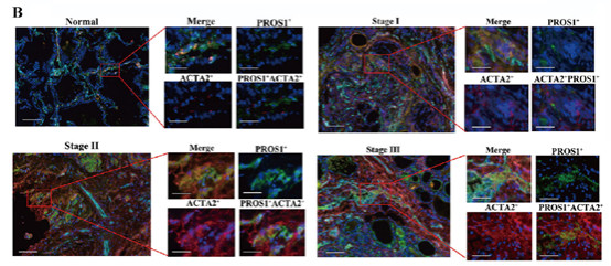
* Results: In normal tissues and non-progressive PTMC, there was almost no colocalization between ACTA2+ fibroblasts and PROS1; while in progressive PTMC (Stage II) and PTC (Stage III), more than 80% of ACTA2+ fibroblasts simultaneously expressed PROS1 (red-green signal overlap rate > 75%), directly confirming that adi-CAFs are the main secretory cells of PROS1.
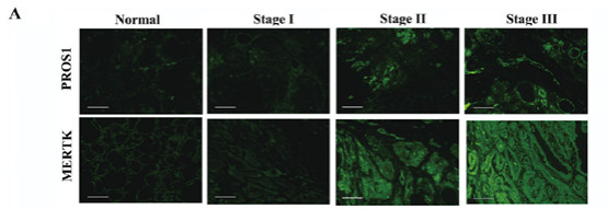
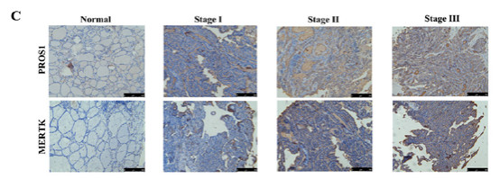
In addition, this kit was also used to verify the tissue expression differences between PROS1 and MERTK: in the tumor area of progressive tissues, the fluorescence signal intensity of PROS1 and MERTK was 3-5 times higher than that in normal tissues, further confirming the correlation between the activation of this axis and disease progression.
V. Summary and Outlook: A "Bridge" from Basic Research to Clinical Translation
This study not only reveals for the first time the core role of the PROS1-MERTK axis in PTMC progression, providing "MERTK" as a potential biomarker and therapeutic target for clinical practice, but also demonstrates the powerful value of the research paradigm of "single-cell + spatial transcriptomics + multiplex fluorescence validation" in the field of cancer microenvironment.
As a key validation tool, Absin's Multiplex IHC/IF Kit (abs50014), with its advantages of high specificity and multi-channel compatibility, provides reliable support for cross-scale mechanism elucidation of "protein-cell-tissue". In the future, Absin will continue to launch higher-quality scientific research tools to facilitate more research breakthroughs in the fields of cancer microenvironment and precision medicine, and provide solid technical support for clinical translation.
(Note: For the screenshot of Figure 5B in the original article and product application details, please refer to the original article in Advanced Science 2025, Volume 12, e13474, or contact the Absin technical team to obtain the complete application protocol.)
VI. Recommendation of Absin Multiplex Fluorescence Immunohistochemistry Kits
| Catalog | Product Name | Specification |
| abs50086 | Absin 2-Color IHC Kit (Anti-Rabbit Secondary Antibody) | 100T |
| abs50087 | Absin 2-Color IHC Kit (Anti-Rabbit and Mouse Secondary Antibody) | 100T |
| abs50088 | Absin 3-Color IHC Kit (Anti-Rabbit Secondary Antibody) | 100T |
| abs50089 | Absin 3-Color IHC Kit (Anti-Rabbit and Mouse Secondary Antibody) | 100T |
| abs50103 | Absin 3-Color IHC Kit B (Anti-Rabbit Secondary Antibody) | 100T |
| abs50104 | Absin 3-Color IHC Kit B (Anti-Rabbit and Mouse Secondary Antibody) | 100T |
| abs50012 | Absin 4-Color IHC Kit (Anti-Rabbit and Mouse Secondary Antibody) | 20T/50T/100T |
| abs50028 | Absin 4-Color IHC Kit(Anti-Rabbit Secondary Antibody) | 20T/50T/100T |
| abs50167 | Absin 4-Color IHC Kit B (Anti-Rabbit and Mouse Secondary Antibody) | 20T/50T/100T |
| abs50168 | Absin 4-Color IHC Kit (Anti-Rabbit Secondary Antibody) | 20T/50T/100T |
| abs50013 | Absin 5-Color IHC Kit (Anti-Rabbit and Mouse Secondary Antibody) | 20T/50T/100T |
| abs50029 | Absin 5-Color IHC Kit (Anti-Rabbit Secondary Antibody) | 20T/50T/100T |
| abs50014 | Absin 6-Color IHC Kit (Anti-Rabbit and Mouse Secondary Antibody) | 20T/50T/100T |
| abs50030 | Absin 6-Color IHC Kit (Anti-Rabbit Secondary Antibody) | 20T/50T/100T |
| abs50048 | Absin 6-Color mlHC Kit(plus) (Anti-Rabbit Secondary Antibody) | 20T/50T/100T |
| abs50049 | Absin 6-Color IHC Kit (plus) (Anti-Rabbit and Mouse Secondary Antibody) | 20T/50T/100T |
| abs50015 | Absin 7-Color IHC Kit (Anti-Rabbit and Mouse Secondary Antibody) | 20T/50T/100T |
| abs50031 | Absin 7-Color IHC Kit(Anti-Rabbit Secondary Antibody) | 20T/50T/100T |
| abs50037 | Absin 7-Color IHC Kit (Anti-Rabbit and Mouse Secondary Antibody) | 20T/50T/100T |
| abs50038 | Absin 7-Color IHC Kit (Anti-Rabbit Secondary Antibody) | 20T/50T/100T |
| abs50165 | Absin 7-Color IHC Kit (Anti-Rabbit Secondary Antibody) | 20T/50T/100T |
| abs50166 | Absin 7-Color IHC Kit (Anti-Rabbit&mouse Secondary Antibody) | 20T/50T/100T |
| abs50018 | Absin 10-Color IHC Kit | 100T |
| abs50083 | Lung Cancer Tumor Microenvironment mIHC Detection Kit (I) | 20T |
| abs50084 | Lung Cancer Tumor Microenvironment mIHC Detection Kit (II) | 20T |
Related News
- PTM Analysis is Essential for Antibody Research 7/10/2026
- BioMed X and Boehringer Ingelheim Expand XSeed Labs to Advance Next-Generation E 7/10/2026
- Everest Medicines Announces Close of Exclusive Licensing Agreement with Travere 7/10/2026
- Pan Modification Microspheres for PTM Proteomics 7/9/2026
- Specific Antibodies Drive Targeted Protein Degradation 7/9/2026
- Thermo Fisher Scientific Receives First FBI Approval for Rapid DNA Crime Scene P 7/9/2026
- Selective Chemical Labeling of Protein Lysine Methacrylation 7/8/2026
- Eppendorf Focuses Outreach Programmes to Advance STEM Education in Europe 7/8/2026
- Fumaroylation: A New Hub Linking Metabolism and Epigenetics 7/7/2026
- OECD Publishes Test Guideline for ToxTracker® 7/7/2026