
Home > News > Mouse T Cell Activation: A Systematic Guide to Developmental Differentiation and Immune Function Characterization
Mouse T Cell Activation: A Systematic Guide to Developmental Differentiation and Immune Function Characterization
- 1. Literature Information
Research Focus: Systematic analysis of murine T lymphocyte developmental stages, functional subset differentiation, molecular regulatory mechanisms, and characterization methodologies—with a focus on flow cytometry-based detection and in vitro activation technologies.
Core Innovation: Establishing a comprehensive framework for T cell research, integrating developmental programming, subset classification, functional assessment, and technical optimization to advance understanding of adaptive immunity and support translational applications.
2. Research Background
T lymphocytes serve as the cornerstone of adaptive immunity, executing antigen-specific immune responses through a highly ordered developmental and functional program. Originating from bone marrow progenitors, T cells undergo rigorous maturation in the thymus, followed by differentiation into specialized subsets that orchestrate immune defense, tolerance, and tissue homeostasis. Dissecting T cell developmental stages, subset heterogeneity, and activation dynamics is critical for elucidating immunological mechanisms, diagnosing immune disorders, and developing cell therapies (e.g., CAR-T/TCR-T). However, the complexity of T cell biology—including diverse subset phenotypes, dynamic differentiation pathways, and context-dependent functional states—poses significant challenges to systematic characterization. Traditional research methods often lack standardization, leading to inconsistent results. There is an urgent need for a unified, multi-dimensional approach to analyze T cell development, differentiation, and activation, supported by reliable tools and standardized protocols.
3. Research Approaches
To comprehensively characterize T cell biology, the research team employed an integrated, technology-driven strategy:
Developmental Tracing: Mapping T cell maturation from bone marrow progenitors to thymic subsets (double-negative, intermediate single-positive, double-positive, single-positive) using surface marker profiling (CD44, CD25, CD117, CD4, CD8, CD3).
Subset Classification: Defining functional T cell subsets (Th1/Th2/Th17/Treg) based on transcription factor expression (T-bet, GATA3, RORγt, Foxp3), cytokine secretion, and chemokine receptor signatures.
Flow Cytometry Optimization: Establishing multi-color flow cytometry protocols for precise subset identification, including surface marker staining, intranuclear transcription factor detection, and memory/activation status analysis (CD44, CD62L, CD69, CD25).
Functional Validation: Combining in vitro activation (via PMA/ionomycin or antigen stimulation) with proliferation assays, cytotoxicity tests, and cytokine profiling to link phenotype to function.
Reference Standard Establishment: Determining the composition of healthy mouse peripheral blood T cells to provide a baseline for comparative studies.
4. Research Outcomes
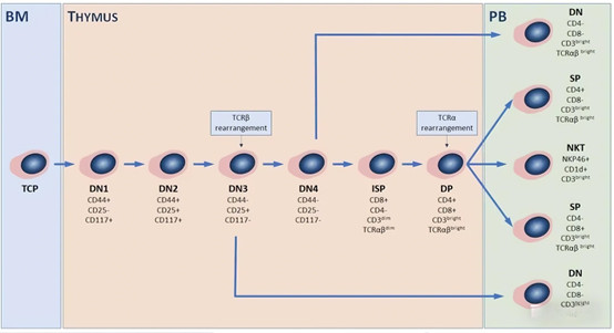
4.1 Key Stages of T Lymphocyte Development
T cells undergo a highly programmed developmental process:
Origin: Bone marrow-derived progenitors migrate to the thymus.
Double-Negative (DN) Stages: DN1-DN4 subphases, defined by CD44, CD25, and CD117 expression; pre-TCR expression initiates at DN3.
Intermediate Single-Positive (ISP) Stage: Transition to CD8+ ISP before acquiring CD4 to become double-positive (DP) cells.
Selection: DP cells undergo positive (MHC recognition) and negative (self-antigen tolerance) selection.
Differentiation: Mature into CD4+ or CD8+ single-positive (SP) αβT cells; alternative pathways produce γδT cells and NKT cells.
4.2 Functional T Cell Subsets and Molecular Regulation
Three principal functional subsets are defined by phenotype, transcription factors, and cytokines:
Helper T Cells (Th): CD4+ subsets including Th1 (T-bet+, IFN-γ+/TNF-α+, CXCR3/CCR5+), Th2 (GATA3+, IL-4/5/13+, CCR3/CCR4+), Th17 (RORγt+, IL-17/22+, CCR6/IL-23R+), and Th22 (aryl hydrocarbon receptor+, IL-22+).
Cytotoxic T Cells (Tc): CD8+, MHC class I-restricted, eliminating targets via perforin-granzyme and Fas/FasL pathways.
Regulatory T Cells (Treg): Foxp3+, CD25+/CTLA-4+, secreting IL-10/TGF-β to maintain immune tolerance.
4.3 Flow Cytometry for Precise T Cell Characterization
Multicolor flow cytometry enables comprehensive profiling through a stepwise approach:
Basic Gating: CD45+CD3+ to isolate total T cells, followed by CD4/CD8 subdivision.
Transcription Factor Detection: Intranuclear staining for T-bet, GATA3, RORγt, and Foxp3.
Memory Status: CD44/CD62L combinations distinguish naive (CD44loCD62Lhi), central memory (CD44hiCD62Lhi), and effector memory (CD44hiCD62Llo) subsets.
Activation Markers: CD69 (early activation) and CD25 (sustained activation) reflect functional status.
Critical technical considerations: Antibody titration, standardized fixation/permeabilization, and sufficient event acquisition for rare subsets.
4.4 T Cell Composition in Healthy Mouse Peripheral Blood
Physiological T cell distribution is stable:
CD4+ Th cells: ~62.8%
CD8+ Tc cells: ~34.53%
Double-negative cells (primarily γδT cells): ~2.56%
Double-positive cells: ~0.12%
Memory Phenotype: ~70% of CD4+/CD8+ T cells are naive (CD44loCD62Lhi); remaining are effector memory or central memory subsets.
4.5 Technical Optimization for T Cell Activation Studies
Key considerations for reliable functional research:
Stimulation: PMA/ionomycin or antigen pre-stimulation for intracellular cytokine detection.
Multiparameter Analysis: Combine surface markers, transcription factors, and cytokines for holistic characterization.
Kinetic Monitoring: Track activation marker expression over time to capture dynamic responses.
Functional Validation: Complement phenotypic data with proliferation (e.g., CFSE labeling) and cytotoxicity assays.
Controls: Include unstimulated cells, fluorescence compensation controls, and isotype controls to ensure specificity.
5. Product Empowerment by ANT BIO PTE. LTD.
ANT BIO PTE. LTD.’s UA brand, a leader in recombinant proteins and cell therapy tools, provides a cornerstone product for T cell research: the "CellXViva Mouse T Cell Activation Kit" (Catalog No.: UA090032). This optimized kit is engineered to support the systematic analysis of T cell activation, differentiation, and function, directly enabling the research outcomes outlined above.
Key Roles of the Product in Research:
Standardized Activation: The kit’s anti-CD3ε/anti-CD28 antibody cocktail, combined with specialized culture additives, delivers consistent, efficient in vitro T cell activation—ensuring reproducible downstream functional assays (cytokine secretion, proliferation, cytotoxicity).
High-Quality Cell Preservation: By maintaining superior cell viability and minimizing activation-induced cell death, the kit provides high-purity CD4+/CD8+ T cells for flow cytometry-based subset analysis and transcription factor detection.
Versatile Application Support: Ideal for validating T cell subset differentiation (e.g., Th1/Th17/Treg induction), antigen-specific T cell expansion, and drug screening—supporting both basic immunology and preclinical cell therapy research.
Batch Consistency: Rigorous quality control (sterility, endotoxin, functional validation) ensures uniform performance across experiments, critical for long-term studies and data comparability.
Compatible with flow cytometry, cytokine ELISAs, and proliferation assays, the kit is an indispensable tool for bridging T cell phenotype and function.
6. Brand Mission
ANT BIO PTE. LTD. is dedicated to advancing global life science research through three specialized sub-brands: ABSIN (general reagents, ELISA kits), STARTER (antibodies), and UA (recombinant proteins, cell therapy tools). Leveraging advanced development platforms—including recombinant monoclonal antibody generation, rapid antibody development, recombinant protein expression (E.coli, CHO, HEK293, Insect Cells), One-Step ELISA, and PTM Pan-Modification Antibody platforms—we deliver high-quality, compliant products certified by EU 98/79/EC, ISO9001, and ISO13485. Our mission is to partner with biopharmaceutical companies, research institutions, and scientists worldwide, providing innovative, reliable reagents and solutions that accelerate discoveries in immunology, cell therapy, and precision medicine.
7. Related Product List
| UA090032 | Mouse T Cell Activation Kit / CellXViva Mouse T cell Activation Kit | Host : Mouse |
8. AI Disclaimer
This article is AI-compiled and interpreted based on the original work. All intellectual property (e.g., images, data) of the original publication shall belong to the journal and the research team. For any infringement, please contact us promptly and we will take immediate action.
ANT BIO PTE. LTD. – Empowering Scientific Breakthroughs
At ANTBIO, we are committed to advancing life science research through high-quality, reliable reagents and comprehensive solutions. Our specialized sub-brands (Absin, Starter, UA) cover a full spectrum of research needs, from general reagents and kits to antibodies and recombinant proteins. With a focus on innovation, quality, and customer-centricity, we strive to be your trusted partner in unlocking scientific mysteries and driving medical progress. Explore our product portfolio today and elevate your research to new heights.
Related News
- Thrombin Antibodies: How Aptamer Technology Revolutionizes Anticoagulation Treat 6/15/2026
- Monoclonal Antibodies: Achieving Precise Targeted Therapy Through Structural Opt 6/14/2026
- Monoclonal Antibodies: Achieving Precise Targeted Therapy Through Structural Opt 6/13/2026
- The Application and Validation of Serine/Threonine Phosphorylation Antibodies in 6/12/2026
- Hua Medicine Advances Glucose Homeostasis Platform at ADA 2026, Showcasing Dorza 6/12/2026
- How Do Phosphorylated Tau Antibodies Reveal the Mechanism of Protective Mutation 6/11/2026
- Everest Medicines Secures Exclusive License for Sumecigrel in Asia-Pacific, Expa 6/11/2026
- Merck Appoints New Heads of Discovery Solutions and Strategy & BD for Life Scien 6/9/2026
- AOP Health Targets Challenging Blood Cancers with First-In-Class Therapy 6/8/2026
- CD182 Antibody: Analyzing the Multiple Functions of CXCR2 Receptor in the Tumor 6/8/2026