
Home > News > SP263: A Key Tool in Immunohistochemical Labeling
SP263: A Key Tool in Immunohistochemical Labeling
Molecular Mechanism and Characteristics of SP263
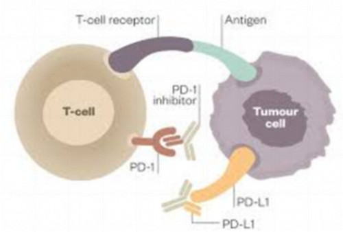
SP263 is a monoclonal antibody targeting PD-L1 (Programmed Death Ligand-1). PD-L1 is a transmembrane protein that belongs to the immune checkpoint molecule family and can suppress the activity of the immune system, particularly the function of T cells. In the tumor microenvironment, PD-L1 binds to PD-1 (Programmed Death Receptor-1), inhibiting T cell activity and helping tumors escape immune surveillance. This mechanism is a key factor in tumor immune evasion and a target for current immune checkpoint inhibitor therapies.As an antibody specifically recognizing PD-L1, SP263 can bind to PD-L1 molecules with high specificity, enabling the precise detection of PD-L1 expression levels in tumor samples during immunohistochemistry experiments. SP263 has exceptionally high sensitivity and specificity for detecting PD-L1, making it an important tool in cancer immunotherapy, especially in evaluating the efficacy of immune treatments. SP263 is a widely used immunohistochemical marker with significant clinical and experimental research value. It is primarily used to detect the expression of specific molecules in tissue samples, especially in cancer diagnosis and immune therapy. As a monoclonal antibody, SP263 has high specificity and sensitivity, effectively recognizing and labeling target antigens. Particularly in cancer immunotherapy, SP263 is used to assess PD-L1 expression levels, providing essential information for patients' treatment plans.
Mechanism of SP263 Binding to PD-L1
PD-L1 molecules are present on the surface of various immune cells and tumor cells, and their expression levels vary significantly across different types of tumors. SP263 helps researchers or clinicians accurately assess PD-L1 expression levels in tumor tissues by specifically recognizing and binding to the surface region of PD-L1. SP263 can directly bind to the antigen epitope on the PD-L1 molecule, forming a stable antigen-antibody complex, which can then be visualized through immunohistochemical reactions. High expression of PD-L1 is often associated with the immune evasion mechanism of tumors. In various tumor types, such as non-small cell lung cancer, melanoma, and gastric cancer, the overexpression of PD-L1 can lead to a reduced ability of the immune system to recognize and eliminate tumor cells. Therefore, as a reliable detection tool, SP263 provides crucial information for selecting and evaluating immune treatment plans.
Application of SP263 in Immunohistochemistry
Immunohistochemistry (IHC) is a commonly used experimental technique in clinical pathology to detect specific proteins or antigens in tissue samples. By using specific antibodies, immunohistochemical techniques help doctors or researchers identify and locate specific molecules inside or outside of cells, providing strong support for disease diagnosis and treatment. SP263, as a specific antibody for PD-L1, is widely used in IHC testing to evaluate PD-L1 expression in tumor tissues.
The main application of SP263 in immunotherapy lies in the detection of PD-L1 expression in cancer patients. By performing immunohistochemical staining on tumor tissue samples, SP263 helps doctors assess the level of PD-L1 expression, providing a reference for the use of immune checkpoint inhibitors. In many types of tumors, patients with higher PD-L1 expression levels tend to respond better to immune checkpoint inhibitor treatments. Therefore, using SP263 to assess PD-L1 expression can assist doctors in selecting the most appropriate immune therapy strategy. For example, in non-small cell lung cancer (NSCLC) patients, the level of PD-L1 expression is closely related to the efficacy of immune therapy. Research has found that patients with high PD-L1 expression respond better to PD-1/PD-L1 inhibitors, while patients with low PD-L1 expression may need alternative treatments. Therefore, SP263 plays a crucial role in PD-L1 evaluation for these patients, helping doctors optimize treatment plans.
Application of SP263 in Tumor Diagnosis
In tumor diagnosis, evaluating PD-L1 expression levels in the tumor microenvironment has become an important biomarker, particularly in the context of immunotherapy. Immune checkpoint inhibitors (such as PD-1/PD-L1 inhibitors) have been proven to significantly improve the treatment outcomes of certain types of cancer patients. Therefore, detecting PD-L1 expression levels helps doctors determine which patients may benefit from immunotherapy, becoming an important task in cancer treatment.
SP263 is widely used in the immunohistochemical detection of clinical samples, especially in assessing PD-L1 expression levels in tumors such as lung cancer, melanoma, and head and neck cancer. SP263 can make PD-L1 molecules in tumor tissues visible through staining reactions, helping pathologists precisely determine the immune evasion mechanisms of tumors.
In certain tumor types, PD-L1 expression may be closely related to clinical and pathological characteristics. Studies have shown that tumors with higher PD-L1 expression often exhibit stronger immune evasion abilities and may be associated with worse prognosis. Therefore, using SP263 for PD-L1 expression detection not only helps in diagnosing tumors but also provides important evidence for treatment decisions.
Future Development and Clinical Prospects of SP263
With the continuous progress of immunotherapy, the importance of SP263 as a PD-L1 detection tool is becoming increasingly prominent. In the future, SP263 is expected to play a key role not only in cancer immunotherapy but also in other fields, such as autoimmune diseases and infectious diseases. Especially in the field of cancer immunotherapy, with the introduction of new immune checkpoint inhibitors, SP263 may become part of standardized testing protocols, further improving the accuracy and efficacy of immunotherapy.
Click on the product catalog numbers below to access detailed information on our official website.
Product Information
| S0B0038 | Invivo anti-mouse PD-L1 Recombinant mAb | Host : Rat |
| UA010003 | PD-L1 Fc Chimera Protein, Human | Host : Human Expression System: HEK293 Conjugation: Unconjugated |
| UA010001 | PD-L1 His Tag Protein, Human | Host : Human Expression System: HEK293 Conjugation: Unconjugated |
| S0B2068 | S-RMab® PD-L1 Recombinant Rabbit mAb (SDT-119-16) |
Host : Rabbit |
| S0B1688 | PD-L1 Recombinant Rabbit mAb (Alexa Fluor® 700 Conjugate) (SDT-119-16) |
Host : Rabbit Conjugation : Alexa Fluor® 700 |
| S0C2014 | PD-L1, CK-Pan, CD8α, CD4, CD163 Antibody Panel | Conjugation: Unconjugated |
| S0C2030 | PD-L1, Regulatory T cell, Cytotoxic T cell and Macrophage Marker (PD-L1, FOXP3, CD8α, CD68, CD163) Antibody Panel |
Conjugation: Unconjugated |
| S0B5429 | Alexa Fluor® 647 Rat Anti-Mouse CD274/B7-H1/PD-L1 Antibody (S-R072) | Host : Rat Conjugation : Alexa Fluor® 647 |
| S0B0593 | Invivo anti-mouse PD-L1 Recombinant mAb (D265A) | Host : Mouse Conjugation: Unconjugated |
| S0B2287P | S-RMab® PD-L1 Recombinant Rabbit mAb,PBS Only (SDT-119-286) | Host : Rabbit Conjugation: Unconjugated |
Related News
- Thrombin Antibodies: How Aptamer Technology Revolutionizes Anticoagulation Treat 6/15/2026
- Monoclonal Antibodies: Achieving Precise Targeted Therapy Through Structural Opt 6/14/2026
- Monoclonal Antibodies: Achieving Precise Targeted Therapy Through Structural Opt 6/13/2026
- The Application and Validation of Serine/Threonine Phosphorylation Antibodies in 6/12/2026
- Hua Medicine Advances Glucose Homeostasis Platform at ADA 2026, Showcasing Dorza 6/12/2026
- How Do Phosphorylated Tau Antibodies Reveal the Mechanism of Protective Mutation 6/11/2026
- Everest Medicines Secures Exclusive License for Sumecigrel in Asia-Pacific, Expa 6/11/2026
- Merck Appoints New Heads of Discovery Solutions and Strategy & BD for Life Scien 6/9/2026
- AOP Health Targets Challenging Blood Cancers with First-In-Class Therapy 6/8/2026
- CD182 Antibody: Analyzing the Multiple Functions of CXCR2 Receptor in the Tumor 6/8/2026